. 2011 Jan;60(1):280-7.
doi: 10.2337/db10-0892. Epub 2010 Oct 15.

miR-200a Prevents renal fibrogenesis through repression of TGF-β2 expression

Affiliations

miR-200a Prevents renal fibrogenesis through repression of TGF-β2 expression

Bo Wang et al. Diabetes. 2011 Jan.

Abstract

Objective: Progressive fibrosis in the diabetic kidney is driven and sustained by a diverse range of profibrotic factors. This study examines the critical role of microRNAs (miRNAs) in the regulation of the key fibrotic mediators, TGF-β1 and TGF-β2.

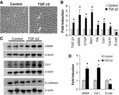
Research design and methods: Rat proximal-tubular epithelial cells (NRK52E) were treated with TGF-β1 and TGF-β2 for 3 days, and expression of markers of epithelial-to-mesenchymal transition (EMT) and fibrogenesis were assessed by RT-PCR and Western blotting. The expression of miR-141 and miR-200a was also assessed, as was their role as translational repressors of TGF-β signaling. Finally, these pathways were explored in two different mouse models, representing early and advanced diabetic nephropathy.

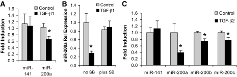
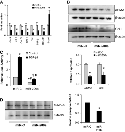
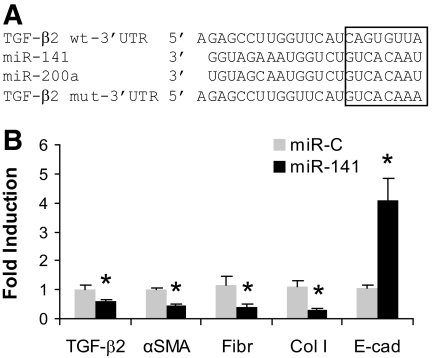
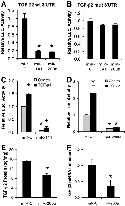
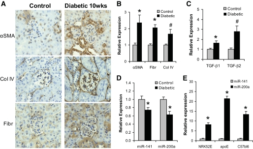
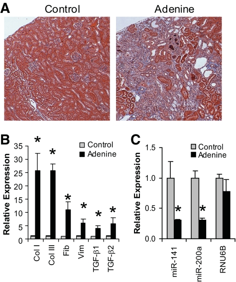
Results: Both TGF-β1 and TGF-β2 induced EMT and fibrogenesis in NRK52E cells. TGF-β1 and TGF-β2 also downregulated expression of miR-200a. The importance of these changes was demonstrated by the finding that ectopic expression miR-200a downregulated smad-3 activity and the expression of matrix proteins and prevented TGF-β-dependent EMT. miR-200a also downregulated the expression of TGF-β2, via direct interaction with the 3' untranslated region of TGF-β2. The renal expression of miR-141 and miR-200a was also reduced in mouse models representing early and advanced kidney disease.

Conclusions: miR-200a and miR-141 significantly impact on the development and progression of TGF-β-dependent EMT and fibrosis in vitro and in vivo. These miRNAs appear to be intricately involved in fibrogenesis, both as downstream mediators of TGF-β signaling and as components of feedback regulation, and as such represent important new targets for the prevention of progressive kidney disease in the context of diabetes.


Figures

FIG. 1.
FIG. 1.
TGF-β2 induces EMT-like changes in proximal tubular epithelial cells. A: NRK52E cells were cultured in the presence of TGF-β2 (10 ng/ml, 3 days). Light microscopy images (×20) demonstrate that TGF-β2 causes a loss of the typical epithelial morphology to larger and more irregular shaped cells typical of the myofibroblast phenotype. B: After treatment with TGF-β2 (10 ng/ml, 3 days), gene expression levels were assessed by real-time QPCR. A significant increase was observed in the expression of profibrotic factors (TGF-β1, CTGF), fibrotic genes (αSMA, fibronectin [Fib], collagen [Col] I and IV), and PAI-1, but E-cadherin (E-cad) was significantly reduced (*P < 0.05 compared with control). C: Western analysis demonstrated that αSMA and Col I were both significantly elevated at the protein level after TGF-β2 treatment, while E-cadherin was decreased. D: The results from the Western analysis in C are shown as a graph (*P < 0.05 compared with control).
FIG. 2.
FIG. 2.
TGF-β1-induced ECM gene and TGF-β2 expression changes in proximal tubular epithelial cells. A: NRK52E cells (Dulbecco's modified Eagle medium, 25 mmol/l glucose, 2% serum) were treated with TGF-β1 (10 ng/ml, 3 days), and the expression of several genes was assessed by real-time QPCR. Significant changes are indicated (*P < 0.05 compared with control). B: The change in expression of TGF-β1 and TGF-β2 genes was assessed by real-time QPCR, and significant changes are indicated (*P < 0.0005 compared with control). C: TGF-β2 proteins levels were measured by ELISA and expressed as pg/mg (*P < 0.0005 compared with control). D: NRK52E cells were incubated with either control IgG (1 μg/ml) or TGF-β2-specific neutralizing antibody (1 μg/ml) for 1 h, and then TGF-β1 was added (10 ng/ml). Cells were harvested 3 days later and subjected to real-time QPCR analysis. The TGF-β2 antibody prevented the increased expression of αSMA, Collagen I, and fibronectin that is induced by TGF-β1, compared with the IgG control antibody (*P < 0.05 compared with control). E: Cells were then treated with TGF-β1 (10 ng/ml, 3 days) in the presence or absence of SB432542, the TGF-β2 receptor inhibitor. Real-time QPCR analysis confirmed that the gene expression changes induced by TGF-β1 are attenuated by SB432542 (*P < 0.05 and #P < 0.001 compared with control).
FIG. 3.
FIG. 3.
TGF-β1-induced changes in miRNA expression. A: NRK52E cells were cultured in the presence of TGF-β1 (10 ng/ml, 3 days) before miRNA expression levels were assessed by real-time QPCR. A significant decrease was observed in miR-200a but not in miR-141 (*P < 0.03 compared with control). B: SB432542 also attenuated the TGF-β1-induced reduction of miR-200a (*P < 0.02 compared with control). C: Treatment of NRK52E cells with TGF-β2 (10 ng/ml, 3 days) also caused reduction in the expression of miR-200a, miR-200b, and miR-200c (*P < 0.05 compared with miR-control).
FIG. 4.
FIG. 4.
miR-200a represses the expression of ECM proteins. A: NRK52E cells were transfected with miR-200a (100 nmol/l), and RNA was harvested after 3 days for real-time QPCR analysis. miR-200a resulted in significantly decreased expression of several ECM proteins including αSMA, collagen (Col) I and IV, and fibronectin (Fibr) (*P < 0.05 compared with control transfected cells). Expression of TGF-β2 was also significantly decreased as was PAI-1, which is downstream of the TGF-β signaling pathway. The expression of E-cadherin (E-cad) was significantly elevated. B: Western analysis demonstrated a significant decrease in αSMA and collagen I by miR-200a, consistent with the RNA expression analysis (*P < 0.05 compared with control transfected cells). The Westerns were quantified and also shown in graph format below the Western blots (*P < 0.05 compared with control). C: NRK52E cells were cotransfected with the p(CAGA)12 SMAD3 activity reporter construct, a β-galactosidase construct, and miR-200a. Four h later, the cells were treated with TGF-β1, and cells were harvested after 3 days. TGF-β1 resulted in increased SMAD3 activity with miR-C, which was strongly inhibited by miR-200a (*P < 0.00005 compared with miR-C control; #P < 0.0005 compared with miR-C with TGF-β1; $P < 0.002 compared with miR-200a control). D: Western analysis of phospho-SMAD3 and total SMAD3 levels in miR-200a-transfected NRK52E demonstrating reduced SMAD3 phosphorylation relative to total SMAD3 protein (*P < 0.05 compared with miR-C control). The Westerns were quantified and shown in graph format (*P < 0.05 compared with miR-C).
FIG. 5.
FIG. 5.
miR-141a shares the same seed sequence with miR-200a and represses the expression of ECM proteins. A: Alignment of the miR-141/200a sequences and the targeted area of the 3′UTR of TGF-β2 (http://www.targetscan.org). Also shown is the altered sequence of the mutant 3′UTR of TGF-β2. B: Proximal tubular cells were transfected with either miR-control or miR-141 (100 nmol/l), and the expression of certain genes was assessed by real-time QPCR. As with miR-200a, miR-141 was able to significantly reduce the expression of αSMA, fibronectin (Fibr), and collagen I (Col I) and resulted in increased expression of E-cadherin (E-cad) (*P < 0.05 compared with control). The expression of TGF-β2 was also significantly reduced.
FIG. 6.
FIG. 6.
The TGF-β2 3′UTR is regulated by miR-141/200a. A: NRK52E cells were transfected with TGF-β2 3′UTR luciferase reporter plasmid (1 μg), β-galactosidase plasmid (0.2 μg), and either miR-control (miR-C), miR-141, or miR-200a (100 nmol/l), and cells were analyzed for β-galactosidase and luciferase activity after 3 days. Both miR-141 and miR-200a were able to significantly repress luciferase activity from the TGF-β2 3′UTR (*P < 0.05 compared with control transfected cells). B: No activity of miR-141 and miR-200a against the mutant TGF-β2 3′UTR was observed. C: miR-141 and (D) miR-200a were able to prevent the increased luciferase activity induced by TGF-β1 on the TGF-β2 3′UTR. E: Total TGF-β2 was significantly decreased at the protein level as measured by ELISA and (F) at the mRNA level as measured by QPCR in NRK52E cells, 3 days after transfection with miR-200a (*P < 0.05, compared with control).
FIG. 7.
FIG. 7.
Changes in gene and miR-200a expression in diabetic mouse kidney cortex. A: Immunohistochemical analysis demonstrated increased expression for αSMA, collagen IV (Col IV), and fibronectin (Fibr) in the diabetic mouse kidney cortex compared with control. B: mRNA was extracted from the renal cortex of control and 10-week diabetic apoE mice (n = 8 per group). Gene expression was assessed by real-time QPCR for a number of genes, revealing significantly increased expression of αSMA, fibronectin, and collagen IV at the RNA level (*P < 0.01 and #P < 0.05 compared with control). C: Expressions of TGF-β1 and TGF-β2 were also elevated at the mRNA level in diabetic mouse kidney cortex (*P < 0.05 and #P < 0.01 compared with control). D: The increased expressions of TGF-β1 and TGF-β2 were associated with decreased expressions of miR-141 and miR-200a in diabetic kidney (*P < 0.05 compared with control). E: Relative expression levels of miR-141 and miR-200a in NRK52E cells and mouse kidney cortex. (*P < 0.005 compared with control). (A high-quality color representation of this figure is available in the online issue.)
FIG. 8.
FIG. 8.
Changes in miR-141/200a expression in the kidney in the adenine-induced renal fibrosis model. A: Trichrome staining of tissue sections from renal cortex from control and adenine-fed C57bl6 mice after 4 weeks of treatment. Blue staining indicates high levels of collagen in the adenine-fed mouse kidney compared with control. B: mRNA was extracted from the renal cortex of control and adenine-fed C57bl6 mice (n = 4 per group). Gene expression was assessed by real-time QPCR, revealing significantly increased expression of collagen I, collagen III, fibronectin, vimentin, TGF-β1, and TGF-β2. C: The increased expression of TGF-β1, TGF-β2, and collagen was associated with decreased expression of miR-141 and miR-200a but not the appropriate control, RNU6B, in kidney cortex (*P < 0.05 compared with control). (A high-quality color representation of this figure is available in the online issue.)

References

    1. Gregory PA, Bracken CP, Bert AG, Goodall GJ: MicroRNAs as regulators of epithelial-mesenchymal transition. Cell Cycle 2008;7:3112–3118 - PubMed
    1. Jiang X, Tsitsiou E, Herrick SE, Lindsay MA: MicroRNAs and the regulation of fibrosis. FEBS J 2010;277:2015–2021 - PMC - PubMed
    1. Hendrickson DG, Hogan DJ, McCullough HL, Myers JW, Herschlag D, Ferrell JE, Brown PO: Concordant regulation of translation and mRNA abundance for hundreds of targets of a human microRNA. PLoS Biol 2009;7:e1000238. - PMC - PubMed
    1. Burns WC, Twigg SM, Forbes JM, Pete J, Tikellis C, Thallas-Bonke V, Thomas MC, Cooper ME, Kantharidis P: Connective tissue growth factor plays an important role in advanced glycation end product-induced tubular epithelial-to-mesenchymal transition: implications for diabetic renal disease. J Am Soc Nephrol 2006;17:2484–2494 - PubMed
    1. Fan JM, Ng YY, Hill PA, Nikolic-Paterson DJ, Mu W, Atkins RC, Lan HY: Transforming growth factor-beta regulates tubular epithelial-myofibroblast transdifferentiation in vitro. Kidney Int 1999;56:1455–1467 - PubMed

Publication types

MeSH terms