. 2010 Oct 15;24(20):2303-16.
doi: 10.1101/gad.1968210.

CDK12 is a transcription elongation-associated CTD kinase, the metazoan ortholog of yeast Ctk1

Affiliations

CDK12 is a transcription elongation-associated CTD kinase, the metazoan ortholog of yeast Ctk1

Bartlomiej Bartkowiak et al. Genes Dev. .

Abstract

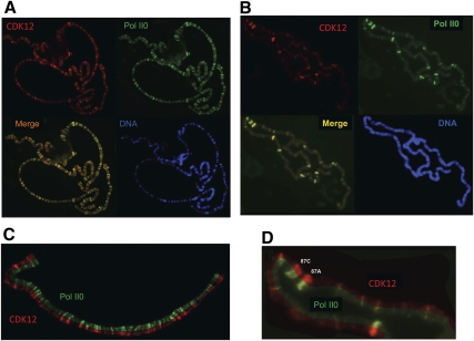
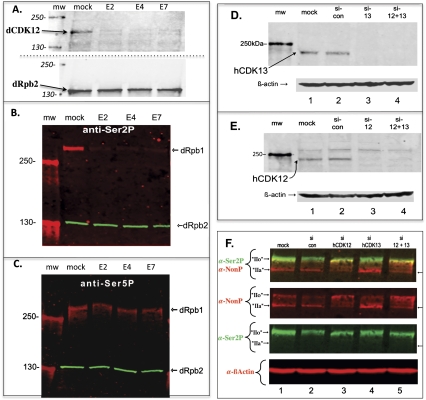
Drosophila contains one (dCDK12) and humans contain two (hCDK12 and hCDK13) proteins that are the closest evolutionary relatives of yeast Ctk1, the catalytic subunit of the major elongation-phase C-terminal repeat domain (CTD) kinase in Saccharomyces cerevisiae, CTDK-I. However, until now, neither CDK12 nor CDK13 has been demonstrated to be a bona fide CTD kinase. Using Drosophila, we demonstrate that dCDK12 (CG7597) is a transcription-associated CTD kinase, the ortholog of yCtk1. Fluorescence microscopy reveals that the distribution of dCDK12 on formaldehyde-fixed polytene chromosomes is virtually identical to that of hyperphosphorylated RNA polymerase II (RNAPII), but is distinct from that of P-TEFb (dCDK9 + dCyclin T). Chromatin immunoprecipitation (ChIP) experiments confirm that dCDK12 is present on the transcribed regions of active Drosophila genes. Compared with P-TEFb, dCDK12 amounts are lower at the 5' end and higher in the middle and at the 3' end of genes (both normalized to RNAPII). Appropriately, Drosophila dCDK12 purified from nuclear extracts manifests CTD kinase activity in vitro. Intriguingly, we find that cyclin K is associated with purified dCDK12, implicating it as the cyclin subunit of this CTD kinase. Most importantly, we demonstrate that RNAi knockdown of dCDK12 in S2 cells alters the phosphorylation state of the CTD, reducing its Ser2 phosphorylation levels. Similarly, in human HeLa cells, we show that hCDK13 purified from nuclear extracts displays CTD kinase activity in vitro, as anticipated. Also, we find that chimeric (yeast/human) versions of Ctk1 containing the kinase homology domains of hCDK12/13 (or hCDK9) are functional in yeast cells (and also in vitro); using this system, we show that a bur1(ts) mutant is rescued more efficiently by a hCDK9 chimera than by a hCDK13 chimera, suggesting the following orthology relationships: Bur1 ↔ CDK9 and Ctk1 ↔ CDK12/13. Finally, we show that siRNA knockdown of hCDK12 in HeLa cells results in alterations in the CTD phosphorylation state. Our findings demonstrate that metazoan CDK12 and CDK13 are CTD kinases, and that CDK12 is orthologous to yeast Ctk1.


Figures

Figure 1.
Figure 1.
Distribution of dCDK12 and hyperphosphorylated RNAPII (Pol II0) on Dm polytene chromosomes, determined by immunofluorescence. (A) Chromosomes from NHS larva reacted with anti-dCDK12 (red), anti-II0 (green), or DAPI (blue: “DNA”). Merge is an overlay of the red and green images. (B) Chromosome set from a heat-shocked larva (20 min at 37°C). (C) Isolated segment of a chromosome arm (non-heat shock) showing split images of dCDK12 (red) and Pol II0 (green). (D) Segment of chromosome 3R showing split images of dCDK12 and Pol II0. Major heat-shock puffs are labeled (87A and 87C).
Figure 2.
Figure 2.
Distribution of dCDK12 and dCyclin T on polytene chromosomes. (A) Chromosomes from NHS larva reacted with anti-dCDK12 (red), anti-dCyclin T (green), or DAPI (blue: “DNA”). The white arrow indicates a developmental puff that stains strongly for CDK12, but weakly for Cyclin T. Red and green dots in the Merge panel indicate bands that are predominantly red (CDK12) or green (Cyclin T), respectively. (B) Chromosome set from a heat-shocked larva (20 min at 37°C) analyzed as for A. Red and green dots in the Merge panel indicate bands that are almost exclusively red (CDK12) or green (Cyclin T), respectively. (C) Isolated chromosome segment (non-heat shock) showing CDK12 (red) and Cyclin T (green). Prominent developmental (ecdysone-induced) puffs 74E and 75B are labeled. Colored dots indicate sites that stain almost uniquely for the corresponding protein. (D) Segment of chromosome 3R (heat shock) with the merged image aligned between images of CDK12 and Cyclin T. Major heat-shock loci are labeled (87A and 87C). Dotted lines facilitate comparisons between red and green images.
Figure 3.
Figure 3.
ChIP analysis of Pol II and dCDK12. (A) ChIP analysis of dCDK12 on Hsp70 using NHS and 10-min heat-shocked (10'HS) samples. (B) ChIP analysis of RNAPII (anti-Rpb3) on Hsp70, as in A. (C) Ratios of dCDK12/RNAPII values on 10-min heat-shocked Hsp70. (D) ChIP analysis of dCDK12 on four constitutively active genes under non-heat-shock conditions. (E) ChIP analysis of RNAPII (anti-Rpb3) on four constitutively active genes under non-heat-shock conditions. (F) Ratios of dCDK12/RNAPII values on four constitutively active genes under non-heat-shock conditions. Measurements in A–F are averages of three biological replicates with standard errors.
Figure 4.
Figure 4.
CTD kinase activities of isolated Drosophila dCDK12 or human hCDK13 enzymes. (A) dCDK12 was eluted from antibody beads using the antigenic peptide, and elution fractions (E1, E2, and E3) were assayed for kinase activity using a βgal-dCTD fusion protein as substrate (apparent molecular weight ∼145 kDa). Coomassie-stained gel of terminated reactions is shown on the left, aligned with the corresponding autoradiogram on the right. (Lanes E1–E3) dCDK12 elutions all show CTD kinase (hyperphosphorylation) activity, as evidenced by the signals in the autoradiogram. No signal is observed in the no-enzyme control lane (fourth lane, no E). (B) dCDK12 was purified using protein A/G beads saturated with affinity-purified anti-dCDK12 IgG. After extensive washing, including a 0.8 M NaCl wash (see the Materials and Methods), the beads were assayed for CTD kinase activity using a GST-yCTD fusion protein as substrate. The Coomassie-stained gel on the left is aligned with the autoradiogram on the right. CTD kinase activity is observed (mobility-shifted band above GST-yCTD position) only when both beads (dCDK12) and CTD substrate are present. (C) hCDK13 was isolated using ion-exchange chromatography followed by immunoaffinity purification (Materials and Methods); the antibody beads were washed extensively and assayed as in B. The mobility-shifted band (autoradiogram) above GST-yCTD position indicates hyperphosphorylation of CTD.
Figure 5.
Figure 5.
dCDK12 and copurifying proteins used for mass spectrometric protein identification. Antibody beads were used to isolate dCDK12 from a nuclear extract. After extensive washing of beads, proteins were solubilized in SDS buffer, subjected to SDS-PAGE, and stained with Coomassie blue. (M) Molecular weight marker lane (values at left); (IP) immunopurified protein lane. Arrow indicates band that is reactive with anti-dCDK12 antibodies. (Lanes a–e) Dotted lines indicate gel slices that were subjected to protein identification analysis in the Duke Proteomics facility.
Figure 6.
Figure 6.
Complementation of bur1 ts allele by chimeric (yeast/human) kinases. (A) The bur1-8 (ts) yeast strain was transformed by empty vector or by vector carrying the 1/9 chimeric kinase construct (cf. Supplemental Fig. S3), and independent transformants were tested for growth at 30°C and 37°C (fivefold serial dilutions spotted in rows). (WT) BUR1+ control strain. (B) The bur1-8 (ts) strain was transformed by empty vector or by vector carrying the 1/13 chimeric kinase construct, and independent transformants were tested for growth at 30°C and 37°C (fivefold serial dilutions spotted in rows). (WT) BUR1+ control strain.
Figure 7.
Figure 7.
RNAi knockdown of dCDK12, hCDK12, or hCDK13 in cultured cells. (A) Mock and RNAi (dsRNA targeting exons E2, E4, or E7 of dCDK12 = CG7597) treatments were carried out for 48 h on cultured Drosophila S2 cells, extracts were prepared, and Western blot analysis was performed using rabbit affinity-purified anti-dCDK12 IgG. Goat affinity-purified anti-dRpb2 IgG was used to assess sample loading. Size standards are in the mw lane, and values in kilodaltons indicated at left. (B) Mock and RNAi treatments and analysis were as in A, except that the phospho-CTD (PCTD) of dRpb1 was detected using mouse anti-Ser2P mAb (H5). Again, dRpb2 was a loading control. (C) Mock and RNAi treatments and analysis were as in B, except that the PCTD of dRpb1 was detected using mouse anti-Ser5P mAb (H14). (D) Mock and siRNA treatments were performed on HeLa R-19 cells, extracts were prepared after 3 d, and Western blot analysis was carried out using rAP anti-hCDK13 IgG. The siRNAs used targeted hCDK13 (si-13), hCDK12 (si-12), or both (si-12 + 13); control siRNAs (si-con) have no homology with any known mammalian genes. The mw lane contains molecular weight markers, and the value is indicated at left. The blot was also reacted with anti-β-actin to show equal loading of extracts. (E) Mock and siRNA treatments were performed as in A, extracts were prepared after 3 d, and Western blot analysis was carried out using rAP anti-hCDK12 IgG and anti-β-actin. (F) Mock and siRNA treatments were performed on HeLa R-19 cells as in A, and Western blot analysis was carried out using rat anti-Ser2P (3E10) and mouse anti-non-phospho CTD (8WG16) mAbs. After analysis of Rpb1, the blot was reprobed with anti-β-actin antibodies. Note that the intensity of IIo in the si-control as detected by anti-Ser2P (lane 2) is usually the same as in the mock treatment (lane 1).

References

    1. Bentley DL 2005. Rules of engagement: Co-transcriptional recruitment of pre-mRNA processing factors. Curr Opin Cell Biol 17: 251–256 - PubMed
    1. Berro R, Pedati C, Kehn-Hall K, Wu W, Klase Z, Even Y, Geneviere AM, Ammosova T, Nekhai S, Kashanchi F 2008. CDK13, a new potential human immunodeficiency virus type 1 inhibitory factor regulating viral mRNA splicing. J Virol 82: 7155–7166 - PMC - PubMed
    1. Boehm AK, Saunders A, Werner J, Lis JT 2003. Transcription factor and polymerase recruitment, modification, and movement on dhsp70 in vivo in the minutes following heat shock. Mol Cell Biol 23: 7628–7637 - PMC - PubMed
    1. Buratowski S 2009. Progression through the RNA polymerase II CTD cycle. Mol Cell 36: 541–546 - PMC - PubMed
    1. Carty SM, Goldstrohm A, Suñe C, Garcia-Blanco MA, Greenleaf AL 2000. Protein-interaction modules that organize nuclear function: FF domains of CA150 bind the phospho-CTD of RNA polymerase II. Proc Natl Acad Sci 97: 9015–9020 - PMC - PubMed

Publication types

MeSH terms