. 2010 Dec;177(6):3061-70.
doi: 10.2353/ajpath.2010.100158. Epub 2010 Oct 15.

The alternative and terminal pathways of complement mediate post-traumatic spinal cord inflammation and injury

Affiliations

The alternative and terminal pathways of complement mediate post-traumatic spinal cord inflammation and injury

Fei Qiao et al. Am J Pathol. 2010 Dec.

Abstract

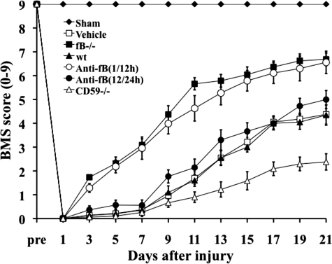
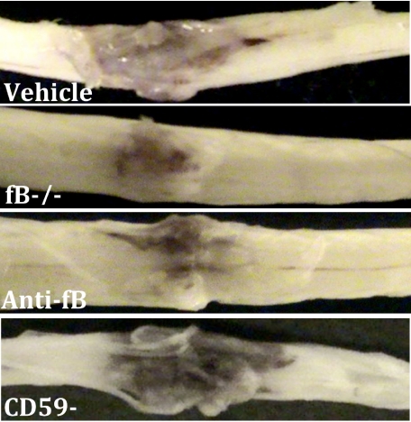
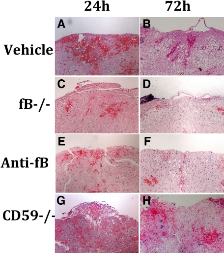
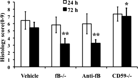
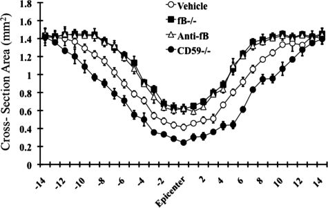
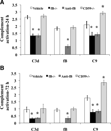
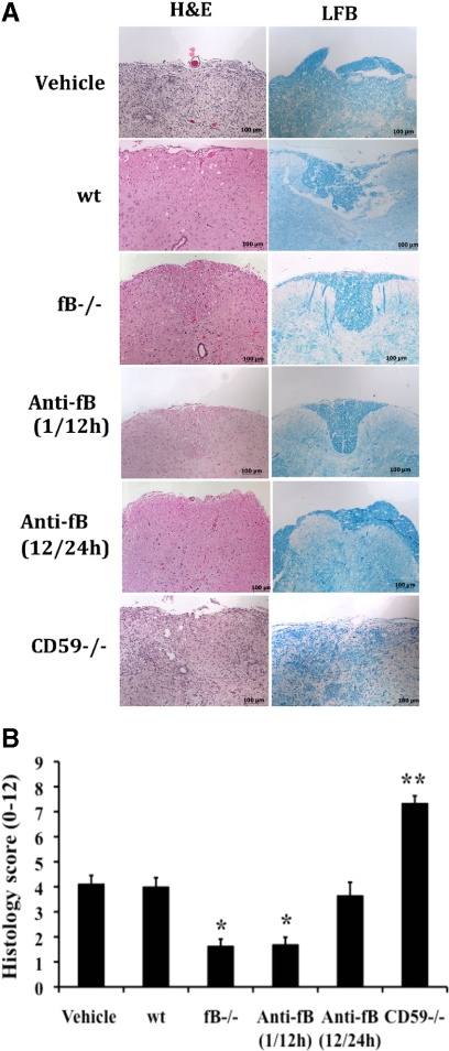
Complement is implicated in the inflammatory response and the secondary neuronal damage that occurs after traumatic spinal cord injury (SCI). Complement can be activated by the classical, lectin, or alternative pathways, all of which share a common terminal pathway that culminates in formation of the cytolytic membrane attack complex (MAC). Here, we investigated the role of the alternative and terminal complement pathways in SCI. Mice deficient in the alternative pathway protein factor B (fB) were protected from traumatic SCI in terms of reduced tissue damage and demyelination, reduced inflammatory cell infiltrate, and improved functional recovery. In a clinically relevant paradigm, treatment of mice with an anti-fB mAb resulted in similarly improved outcomes. These improvements were associated with decreased C3 and fB deposition. On the other hand, deficiency of CD59, an inhibitor of the membrane attack complex, resulted in significantly increased injury and impaired functional recovery compared to wild-type mice. Increased injury in CD59-deficient mice was associated with increased MAC deposition, while levels of C3 and fB were unaffected. These data indicate key roles for the alternative and terminal complement pathways in the pathophysiology of SCI. Considering a previous study demonstrating an important role for the classical pathway in promoting SCI, it is likely that the alternative pathway plays a critical role in amplifying classical pathway initiated complement activation.


Figures

Figure 1
Figure 1
Locomotor recovery after SCI in complement deficient and inhibited mice. Open-field 10-point BMS scores were recorded for 21 days after contusion-induced SCI in the indicated groups of mice. Anti-fB mAb treatment group received an i.v. injection of 2 mg at 1 and 12 or 12 and 24 hours after injury. BMS scores are significantly higher for fB−/− mice and anti-fB mAb (1/12 hours)-treated mice compared to wt or vehicle (PBS)-treated mice from day 3 postinjury. No significant difference between vehicle controls and anti-fB mAb (12/24 hours) treated mice. BMS scores are significantly lower in CD59−/− mice compared to wt and vehicle control mice from day 11 after injury (P < 0. 01). Mean ± SE, n = 8–10.
Figure 2
Figure 2
Macroscopic images of spinal cords isolated at 72 hours postinjury. Mice received anti-fB mAb or PBS (vehicle) at 1 and 12 hours after injury. Representative images shown, n = 6–8 per group.
Figure 3
Figure 3
Histopathology of spinal cord sections after SCI. A–H: Transverse sections from the epicenter of injury were prepared from spinal cords isolated at 24 and 72 hours after injury and stained with H&E. Mice received anti-fB mAb or PBS (vehicle) at 1 and 12 hours after injury. Representative images shown, n = 6 per group.
Figure 4
Figure 4
Quantitative assessment of histopathological inflammation and injury. H&E-stained sections from the epicenter of injury were scored from 0 to 3 for the presence and intensity of inflammatory cell infiltration, neuronal vacuolation, and hemorrhage (0 = no evidence, 3 = severe). Scores were then expressed as a cumulative score of 0–9. Assessments were made from spinal cords isolated at 24 and 72 hours postinjury. Mice received anti-fB mAb or PBS (vehicle) at 1 and 12 hours after injury. *P < 0.01, **P < 0.001 compared to vehicle control. Mean ± SD, n = 6 per group.
Figure 5
Figure 5
Morphometric analysis of tissue sparing 3 days after injury. The cross-sectional area of H&E stained spinal cord sections was measured at 150 μm increments extending 2 mm either side of the injury site. There was a significant level of tissue sparing in fB−/− mice and anti-fB–treated mice compared with vehicle control (PBS)-treated mice, and a significant decrease in tissue sparing in CD59−/− mice compared with wt (at injury site and out to 1.2 mm each side of the injury site, P < 0.01). Mice received anti-fB mAb or PBS at 1 and 12 hours after injury. Mean ± SD n = 6 per group.
Figure 6
Figure 6
Neutrophil (A) and macrophage (B) infiltration after SCI. Sections from the epicenter of injury were prepared from spinal cords isolated at 24 and 72 hours after injury. Mice received anti-fB mAb or PBS (vehicle) at 1 and 12 hours after injury. Neutrophils and macrophages were identified and counted by immunohistochemistry using anti-mouse Gr-1 and Mac-3 antibodies. Results are expressed as the number of neutrophils/macrophages per mm2. Mean ± SD n = 6 per group. *P < 0.01 vs. vehicle control.
Figure 7
Figure 7
Quantitative assessment of complement deposition post-SCI. The deposition of C3 (C3 days), fB, and C9 (MAC) was analyzed at 24 hours (A) and 72 hours (B) after injury in spinal cord sections from the epicenter of injury. Mice received anti-fB mAb or PBS (vehicle) at 1 and 12 hours after injury. Analysis was performed using immunofluorescence microscopy, and sections scored for fluorescence intensity on a 4 point scale (see Materials and Methods). Mean ± SD, n = 6. *P < 0.01.
Figure 8
Figure 8
Histopathology of spinal cord sections 21 days after injury. A: Images of transverse sections from spinal cords at the epicenter of injury. Sections stained with either H&E or LFB. Representative images, n = 6 per group. B: Quantitative assessment of histopathological inflammation, injury, and demyelination. H&E-stained sections were scored on a 0–3 scale for the presence and intensity of inflammatory cell infiltration, neuronal vacuolation, and hemorrhage. LFB-stained sections scored for demyelination (0 = no evidence, 3 = severe). Anti-fB mAb treatment groups received injections at 1 and 12, or 12 and 24 hours after injury. Vehicle control group (PBS) received injections at 1 and 12 hours after injury. Scores were then expressed as a cumulative score of 0–12. Mean ± SD, n = 6 per group. *P < 0.001 and **P < 0.0001 vs. vehicle control and wt.

Comment in

Similar articles

Cited by

References

    1. Fiore C, Inman DM, Hirose S, Noble LJ, Igarashi T, Compagnone NA. Treatment with the neurosteroid dehydroepiandrosterone promotes recovery of motor behavior after moderate contusive spinal cord injury in the mouse. J Neurosci Res. 2004;75:391–400. - PubMed
    1. Tsai EC, Tator CH. Neuroprotection and regeneration strategies for spinal cord repair. Curr Pharm Des. 2005;11:1211–1222. - PubMed
    1. Hall ED, Springer JE. Neuroprotection and acute spinal cord injury: a reappraisal. NeuroRx. 2004;1:80–100. - PMC - PubMed
    1. Rebhun J, Madorsky JG, Glovsky MM. Proteins of the complement system and acute phase reactants in sera of patients with spinal cord injury. Ann Allergy. 1991;66:335–338. - PubMed
    1. Anderson AJ, Robert S, Huang W, Young W, Cotman CW. Activation of complement pathways after contusion-induced spinal cord injury. J Neurotrauma. 2004;21:1831–1846. - PubMed

Publication types

MeSH terms