. 2010 Dec;177(6):3233-43.
doi: 10.2353/ajpath.2010.100059. Epub 2010 Oct 15.

Angiopoietin-2-driven vascular remodeling in airway inflammation

Affiliations

Angiopoietin-2-driven vascular remodeling in airway inflammation

Sebastien P Tabruyn et al. Am J Pathol. 2010 Dec.

Abstract

Vascular remodeling is a feature of chronic inflammation during which capillaries transform into venules that expand the region of the vasculature in which leakage and leukocyte emigration both occur. Recently, we found that angiopoietin/Tie2 receptor signaling drives the transformation of capillaries into venules at an early stage of the sustained inflammatory response in the airways of mice infected with Mycoplasma pulmonis. However, the precise contributions of both angiopoietin-1 (Ang1) and angiopoietin-2 (Ang2) are not clear. In this study, we sought to determine the contribution of Ang2 to this vascular remodeling. Ang2 mRNA expression levels increased and phosphorylated Tie2 immunoreactivity in mucosal blood vessels decreased, indicative of diminished receptor signaling after infection. Selective inhibition of Ang2 throughout the infection by administration of either of two distinct function-blocking antibodies reduced the suppression of Tie2 phosphorylation and decreased the remodeling of mucosal capillaries into venules, the amount of leukocyte influx, and disease severity. These findings are consistent with Ang2 acting as an antagonist of Tie2 receptors and the reduction of Tie2 phosphorylation in endothelial cells rendering the vasculature more responsive to cytokines that promote both vascular remodeling and the consequences of inflammation after M. pulmonis infection. By blocking such changes, Ang2 inhibitors may prove beneficial in the treatment of sustained inflammation in which vascular remodeling, leakage, and leukocyte influx contribute to its pathophysiology.


Figures
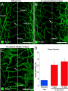
Figure 1
Figure 1
Vascular enlargement after M. pulmonis infection. AC: Confocal microscopic images of blood vessels (green, PECAM-1) in tracheal whole mounts comparing capillaries in pathogen-free mouse (A, arrow) to enlarged, remodeled vessels (arrows) in mice infected with M. pulmonis for 7 days (B) or 14 days (C). D: Measurements of vessels at midpoint of cartilage ring (dotted line) show the magnitude of vascular enlargement after infection. *P < 0.05 compared with pathogen-free. Scale bars = 100 μm.
Figure 2
Figure 2
Increase in Ang2 expression after M. pulmonis infection. A and B: qRT-PCR measurements of expression of Ang1 mRNA (A) and Ang2 mRNA (B) in trachea of pathogen-free mice and mice infected with M. pulmonis for 1, 3, 7, or 14 days. C: Ratio of qRT-PCR measurements of Ang2 mRNA to Ang1 mRNA. *P < 0.05 compared to pathogen-free (day 0). D and E: Confocal micrographs of vessels in tracheal whole mounts showing Ang2 (red) and PECAM-1 (green) immunoreactivities of a capillary in a pathogen-free mouse (D) and a remodeled vessel over a cartilage ring in a mouse infected with M. pulmonis for 7 days (E). Scale bars = 30 μm (D and E).
Figure 3
Figure 3
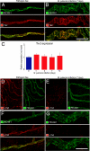
Decrease in Tie2 expression after M. pulmonis infection. A and B: Confocal micrographs showing the similarity of Tie2 (red) in tracheal capillary of pathogen-free mouse (A) and remodeled vessel in mouse infected for 7 days (B). PECAM-1, green. C: qRT-PCR measurements showing no difference in Tie-2 mRNA, expressed relative to β-actin, in trachea of pathogen-free mice (day 0) and mice infected for 1, 3, 7, or 14 days. DG: Confocal micrographs at lower (D and E) and higher (F and G) magnifications showing weaker p-Tie2 immunoreactivity (red) in remodeled vessels after infection for 7 days (E and G) than in tracheal capillary of pathogen-free mouse (D and F). Images were acquired with the same fluorescence intensity setting. Scale bars: 30 μm (A and B); 100 μm (D and E); 30 μm (F and G).
Figure 4
Figure 4
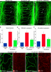
Reduction in vascular enlargement and p-Tie2 suppression by Ang2 blockade with REGN145. A−C: Confocal micrographs comparing the size of blood vessels (green, PECAM-1) in tracheal whole mounts from a pathogen-free mouse (A) and mice infected with M. pulmonis for 7 days with concurrent treatment with normal IgG (B) or REGN145 5 mg/kg (C). Arrows mark blood vessel measurement site over midpoint of cartilage ring. D: Contrasting mean vessel diameters in tracheas of pathogen-free mice and infected mice treated with IgG or REGN145 for 7 days. E and F: qRT-PCR measurements of PECAM-1 (E) and VE-cadherin (F) in tracheas of pathogen-free mice and infected mice treated with IgG or REGN145 for 7 days. *P < 0.05 compared to pathogen-free; #P < 0.05 compared to infected mice treated with IgG. G and H: Pairs of confocal micrographs showing PECAM-1 (green) and p-Tie2 (red) immunoreactivities of tracheal blood vessels after infection for 7 days. p-Tie2 is faint or absent on vessels in infected mouse that received IgG (G) but is stronger on vessels in infected mouse treated with REGN145 (H). Scale bars: 150 μm (AC) or 100 μm (G and H).
Figure 5
Figure 5
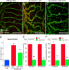
Reduction in adhesion molecule expression by Ang2 blockade with REGN145. A−C: Confocal micrographs of P-selectin immunoreactivity (red) in blood vessels (green, PECAM-1) of tracheal whole mounts. P-selectin immunoreactivity in pathogen-free trachea (A) is absent in capillaries (arrows) and faint in venules (arrowhead). After M. pulmonis infection, P-selectin is strong in remodeled vessels (arrow) of mouse treated with IgG (B) but weak in vessels (arrow) of mouse treated with REGN145 at a dose of 5 mg/kg (C). D: Fractional area of P-selectin immunoreactivity in regions of vessels over cartilage in pathogen-free mice and in infected mice treated concurrently with IgG or REGN145. E and F: qRT-PCR measurements comparing expression of P-selectin (E), and E-selectin (F) mRNA in tracheas of pathogen-free and infected mice treated with IgG or REGN145 for 7 days. *P < 0.05 compared to pathogen-free; **P < 0.05 compared to infected mice treated with IgG. Scale bars = 150 μm.
Figure 6
Figure 6
Reduction in vascular remodeling by Ang2 blockade with mAb 3.19.3. A−C: Confocal micrographs of P-selectin immunoreactivity (red) in blood vessels (green, PECAM-1) of tracheal whole mount of pathogen-free mouse and mice infected with M. pulmonis for 7 days with concurrent treatment with 10 mg/kg of isotype control IgG (B) or mAb 3.19.3 (C). Arrows mark regions where vessel measurements were made. D: Diameter of tracheal blood vessels measured over midpoint of cartilage rings in pathogen-free mice and mice treated with IgG or mAb 3.19.3 during 7-day infection. E and F: qRT-PCR measurements of expression of PECAM-1 and VE-cadherin (E) or P-selectin and E-selectin (F) in tracheas of infected mice treated with IgG or mAb 3.19.3 for 7 days. Values after Ang2 blockade by mAb 3.19.3 were significantly less. *P < 0.05 compared to pathogen-free; **P < 0.05 compared to infected mice treated with IgG. Scale bars = 100 μm.
Figure 7
Figure 7
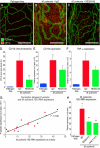
Reduction in leukocyte influx by Ang2 blockade during infection. A−C: Confocal micrographs of leukocytes (red, CD11b) and blood vessels (green, PECAM-1) in regions over cartilage in tracheal whole mount of pathogen-free mouse and mice with M. pulmonis infection for 7 days and concurrent treatment with 5 mg/kg of isotype control IgG (B) or REGN145 (C). D: Fractional area of CD11b immunoreactivity in mucosa over cartilages of pathogen-free mice and infected mice treated with IgG or REGN145. E and F: qRT-PCR measurements of expression of CD11b (E), TNF-α (F) in tracheas of pathogen-free mice and infected mice treated with IgG or REGN145 for 7 days, showing significantly lower values after REGN145 (N = 15). G: Linear regression (r2 = 0.89) of P-selectin expression and M. pulmonis 16S rRNA expression measured by qRT-PCR in tracheas of mice infected for 7 days. H: qRT-PCR measurements of M. pulmonis 16S rRNA expression in tracheas of pathogen-free mice and mice infected and treated for 7 days with control IgG, REGN145, or mAb 3.19.3. *P < 0.05 compared to pathogen-free; **P < 0.05 compared to infected mice treated with IgG. Scale bars = 150 μm.

References

    1. Wilson JW, Hii S. The importance of the airway microvasculature in asthma. Curr Opin Allergy Clin Immunol. 2006;6:51–55. - PubMed
    1. James AL, Pare PD, Hogg JC. The mechanics of airway narrowing in asthma. Am Rev Respir Dis. 1989;139:242–246. - PubMed
    1. Kanazawa H. Role of vascular endothelial growth factor in the pathogenesis of chronic obstructive pulmonary disease. Med Sci Monit. 2007;13:RA189–RA195. - PubMed
    1. de Boer WI, Alagappan VK, Sharma HS. Molecular mechanisms in chronic obstructive pulmonary disease: potential targets for therapy. Cell Biochem Biophys. 2007;47:131–148. - PubMed
    1. McDonald DM. Angiogenesis and remodeling of airway vasculature in chronic inflammation. Am J Respir Crit Care Med. 2001;164:S39–S45. - PubMed

Publication types

MeSH terms