. 2010 Dec;76(24):8093-101.
doi: 10.1128/AEM.01863-10. Epub 2010 Oct 15.

Characterization of the protocatechuate 4,5-cleavage pathway operon in Comamonas sp. strain E6 and discovery of a novel pathway gene

Affiliations

Characterization of the protocatechuate 4,5-cleavage pathway operon in Comamonas sp. strain E6 and discovery of a novel pathway gene

Naofumi Kamimura et al. Appl Environ Microbiol. 2010 Dec.

Abstract

The protocatechuate (PCA) 4,5-cleavage (PCA45) pathway is the essential catabolic route for the degradation of various aromatic acids in the genus Comamonas. All of the PCA45 pathway genes, orf1-pmdKEFDABC, as well as another PCA 4,5-dioxygenase gene, pmdA(II)B(II), were isolated from a phthalate-degrading bacterium, Comamonas sp. strain E6. Disruption of pmdB and pmdD in E6, which code for the β subunit of PCA 4,5-dioxygenase and 2-pyrone-4,6-dicarboxylate (PDC) hydrolase, respectively, resulted in a growth defect on PCA, indicating that these genes are essential for the growth of E6 on PCA. On the other hand, inactivation of pmdB(II) did not affect the growth of E6 on PCA. Disruption of pmdK, which is related to a 4-hydroxybenzoate/PCA transporter of Pseudomonas putida, resulted in growth retardation on PCA. The insertional inactivation of orf1 in E6, whose deduced amino acid sequence has no similarity with proteins of known function, led to the complete loss of growth on PCA and the accumulation of PDC and 4-oxalomesaconate (OMA) from PCA. These results indicated the involvement of orf1 in the PCA45 pathway, and this gene, designated pmdU, was suggested to code for OMA tautomerase. Reverse transcription-PCR analysis suggested that the pmdUKEFDABC genes constitute an operon. The transcription start site of the pmd operon was mapped at 167 nucleotides upstream of the initiation codon of pmdU. The pmd promoter activity was enhanced 20-fold when the cells were grown in the presence of PCA. Inducers of the pmd operon were found to be PCA and PDC, but PDC was the more effective inducer.


Figures

FIG. 1.
FIG. 1.
Proposed catabolic pathway for PCA by Comamonas sp. strain E6 and gene organization of the PCA45 pathway genes. (A) Enzymes: PmdA, small subunit of 4,5-PCD; PmdB, large subunit of 4,5-PCD; PmdC, CHMS dehydrogenase; PmdD, PDC hydrolase; PmdU, putative OMA tautomerase; PmdE, OMA hydratase; PmdF, CHA aldolase/oxaloacetate decarboxylase. Abbreviations: PCA, protocatechuate; CHMS, 4-carboxy-2-hydroxymuconate-6-semialdehyde; PDC, 2-pyrone-4,6-dicarboxylate; OMA, 4-oxalomesaconate; and CHA, 4-carboxy-4-hydroxy-2-oxoadipate. (B) Organization of the pmd gene cluster and pmdAIIBII. Restriction maps of the 9.8-kb SacI fragment carrying the pmd gene cluster and the 4.3-kb KpnI fragment carrying pmdAIIBII are shown. “deletion” and “kan” indicate the positions deleted in EME015 and EME016 and those of kan gene insertions into EME012, EME013, and EME014, respectively. Abbreviations: A, ApaI; B, BglII; Ei, EcoRI; Ev, EcoRV; Hc, HincII; Hd, HindIII; K, KpnI; Ps, PstI; Pv, PvuII; Sa, SacI; Sc, SacII; Sl, SalI; Sp, SphI; St, StuI; Xh, XhoI.
FIG. 2.
FIG. 2.
RT-PCR and qRT-PCR analyses of the PCA45 pathway genes. (A) Agarose gel electrophoresis of RT-PCR products. Total RNA isolated from E6 cells grown in the presence of PCA was used as a template for cDNA synthesis. Lane numbers correspond to the numbers of the amplified regions indicated above. The sizes of molecular weight markers in lane M are indicated on the left. “+” and “−,” with and without reverse transcriptase, respectively. (B) qRT-PCR analysis of the expression of the pmd genes. Total RNA was isolated from E6 cells grown in the presence or absence of PCA. Expression of the pmd genes was measured using a qRT-PCR. Values for each transcript amount were normalized to the level for 16S rRNA and are shown as fold increases over the level for SUC-grown E6 cells, indicated by a dotted line (level of 1.0). The data are mean values ± standard deviations for three independent experiments.
FIG. 3.
FIG. 3.
Disruption of pmdB and pmdBII in Comamonas sp. strain E6. (A) The cells of the pmdB mutant (EME012) were grown in W medium containing 10 mM PCA. Growth levels of E6 and EME012 are indicated by open circles and filled circles, respectively. (B) Complementation of pmdB or pmdBII in EME012 for growth on PCA. Growth levels of E6 cells harboring pJB866 (open circles), EME012 cells harboring pJB866 (filled circles), EME012 cells harboring pJBB1F, which carries pmdB (filled squares), and EME012 cells harboring pJBB2, which carries pmdBII (filled triangles), are shown. The cells were incubated in W medium containing 10 mM PCA, tetracycline, and 3 mM m-toluate. (C) Growth of the cells of E6 (open circles) and pmdBII mutant (EME013; filled circles) in W medium containing 10 mM PCA.
FIG. 4.
FIG. 4.
Growth of pmdD and pmdK mutants on PCA. (A) Growth of the cells of E6 (open circles) and the pmdD mutant (EME016; filled circles) in W medium containing 10 mM PCA. (B) Accumulation of PDC during the incubation of EME016 cells with PCA. The cells of E6 and EME016 were grown in W medium containing 10 mM SUC and PCA. The concentrations of PDC (circles) and PCA (triangles) in the culture incubated with the cells of E6 (open symbols) and EME016 (filled symbols) were monitored using HPLC. (C) Growth of the cells of E6 (open circles) and the pmdK mutant (EME015; filled circles) on PCA under different pH conditions. The E6 and EME015 cells were incubated in W medium containing 10 mM PCA at pH 6.0, 7.0, or 8.0.
FIG. 5.
FIG. 5.
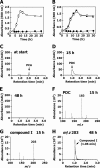
Disruption of orf1 in Comamonas sp. strain E6. (A) Growth of the cells of orf1 mutant (EME014) in W medium containing 10 mM PCA. The growth levels of the cells of E6 and EME014 are indicated by open circles and filled circles, respectively. (B) Complementation of orf1 in the EME014 mutant. The growth levels of E6 cells harboring pJB866 (open circles), EME014 cells harboring pJB866 (filled circles), and EME014 cells harboring pJBEUF, which carries orf1 (filled squares), are shown. The cells were incubated in W medium containing 10 mM PCA, tetracycline, and 3 mM m-toluate. (C to E) Accumulation of PDC and OMA during the degradation of PCA by EME014. The resting cells of EME014 were incubated with 2 mM PCA, and portions of the cultures were collected at the start, at 15 h, and at 48 h. The supernatants of the cultures were analyzed by HPLC. PDC was detected at 315 nm. AU, absorbance units. (F and G) Negative-ion ESI-MS spectra of PDC with a retention time of 1.13 min and compound I with a retention time of 1.06 min, respectively. (H) Extracted ion chromatogram at an m/z of 203 for the supernatants of the culture incubated at 48 h.
FIG. 6.
FIG. 6.
Identification of the pmd promoter region. (A) Deletion analysis of the pmd promoter region. Schematic of the DNA regions fused with lacZ, indicating the location of the deletion, is shown to the left. The β-galactosidase activities of the E6 cells harboring each plasmid grown in the presence or absence of PCA are shown to the right. “+1” indicates the transcription start site of the pmd operon. (B) Primer extension analyses of the pmd operon transcript by use of pEX1. Fluorescent primer extensions were performed using the D4-labeled primer (pEX1) and total RNA isolated from E6 cells harboring pKT10SAF, which carries the entire pmd gene cluster, grown in the presence (left) or absence (right) of PCA. The transcription start site was determined by comparing the retention time of the D4-labeled primer extension product with those of DNA size standards. AU, arbitrary fluorescence units. (C) Nucleotide sequence of the pmd promoter region. The endpoints of deletions are indicated by bent arrows. The inverted repeat sequences (convergent arrows) and the transcription start site (+1) are indicated. The initiation codon of pmdU is double underlined. The horizontal dotted arrow indicates the position of pEX1.

Similar articles

Cited by

References

    1. Abe, T., E. Masai, K. Miyauchi, Y. Katayama, and M. Fukuda. 2005. A tetrahydrofolate-dependent O-demethylase, LigM, is crucial for catabolism of vanillate and syringate in Sphingomonas paucimobilis SYK-6. J. Bacteriol. 187:2030-2037. - PMC - PubMed
    1. Alhapel, A., D. J. Darley, N. Wagener, E. Eckel, N. Elsner, and A. J. Pierik. 2006. Molecular and functional analysis of nicotinate catabolism in Eubacterium barkeri. Proc. Natl. Acad. Sci. U. S. A. 103:12341-12346. - PMC - PubMed
    1. Bagdasarian, M., R. Lurz, B. Rückert, F. C. Franklin, M. M. Bagdasarian, J. Frey, and K. N. Timmis. 1981. Specific-purpose plasmid cloning vectors II. Broad host range, high copy number, RSF1010-derived vectors, and a host-vector system for gene cloning in Pseudomonas. Gene 16:237-247. - PubMed
    1. Blatny, J. M., T. Brautaset, H. C. Winther-Larsen, P. Karunakaran, and S. Valla. 1997. Improved broad-host-range RK2 vectors useful for high and low regulated gene expression levels in gram-negative bacteria. Plasmid 38:35-51. - PubMed
    1. Bolivar, F., and K. Backman. 1979. Plasmids of Escherichia coli as cloning vectors. Methods Enzymol. 68:245-267. - PubMed

Publication types

Associated data

LinkOut - more resources