. 2011 Jan 6;117(1):323-32.
doi: 10.1182/blood-2010-01-261792. Epub 2010 Oct 15.

Opposing roles for HIF-1α and HIF-2α in the regulation of angiogenesis by mononuclear phagocytes

Affiliations

Opposing roles for HIF-1α and HIF-2α in the regulation of angiogenesis by mononuclear phagocytes

Tim D Eubank et al. Blood. .

Abstract

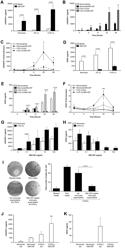
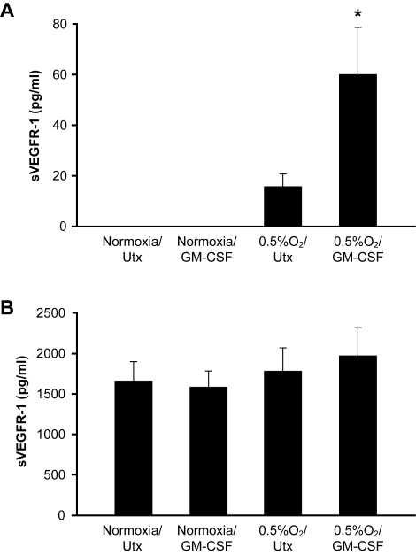
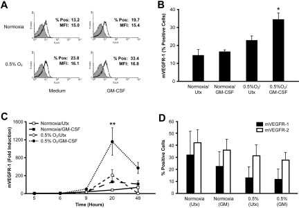
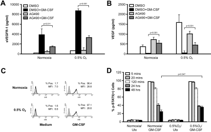
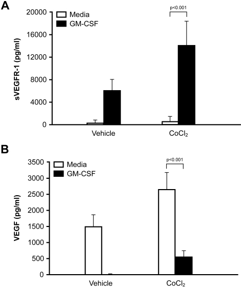
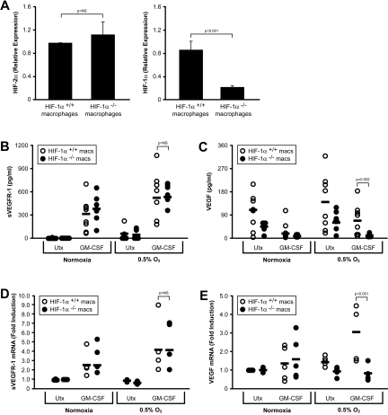
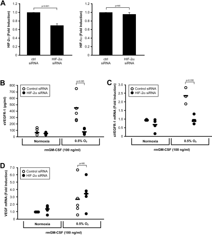
Macrophages contribute to tumor growth through the secretion of the proangiogenic molecule vascular endothelial growth factor (VEGF). We previously observed that monocytes treated with the cytokine granulocyte-macrophage colony-stimulating factor (GM-CSF) produce a soluble form of the VEGF receptor-1 (sVEGFR-1), which neutralizes VEGF biologic activity. The VEGF and VEGFR-1 promoters both contain a hypoxia regulatory element, which binds the hypoxia-inducible factor (HIF) transcription factors under hypoxic conditions. Based on this observation, we examined VEGF and sVEGFR-1 production from monocytes cultured at various O(2) concentrations. The amount of sVEGFR-1 production observed from GM-CSF-treated monocytes increased with decreasing levels of O(2). This sVEGFR-1 was biologically active and sequestered VEGF. To evaluate the role of the HIFs in sVEGFR-1 production, we used macrophages with a genetic deletion of HIF-1α. HIF-1α(-/-) macrophages cultured with GM-CSF at hypoxia secreted diminished amounts of VEGF compared with HIF-1α(+/+) macrophages, whereas sVEGFR-1 secretion was unaffected. In contrast, siRNA-mediated knockdown of HIF-2α inhibited the production of sVEGFR-1 in response to GM-CSF and low O(2), whereas VEGF production was unaffected. These studies suggest that hypoxia, generally thought to promote angiogenesis, can induce antiangiogenic behavior from macrophages within a GM-CSF-rich environment. Furthermore, these results suggest specific and independent roles for HIF-1α and HIF-2α in hypoxic macrophages.


Figures

Figure 1
Figure 1
Production of sVEGFR-1 by mononuclear phagocytes increases with decreasing O2 concentration. (A) Human peripheral blood monocytes were cultured at 21% (ambient) O2, 5% O2 (mild hypoxia), or 0.5% O2 (severe hypoxia) in media alone or media containing 100 ng/mL GM-CSF. After 48 hours, culture supernatants were harvested and analyzed for sVEGFR-1 content by ELISA. Data are mean ± SEM of 7 normal donors. (B) Monocytes were stimulated with GM-CSF at ambient O2 or at 0.5% O2. Culture supernatants were harvested at the indicated time points and analyzed for sVEGFR-1 content by ELISA. Data are the mean ± SEM of 6 normal donors. (C) Monocytes were stimulated as in panel B and analyzed for sVEGFR-1 transcript by real-time PCR at the indicated time points. Data are the mean ± SEM of 6 normal donors. (D) Supernatants from the same cells as in panel A were measured using an ELISA that detects only bioavailable VEGF (ie, VEGF that is not bound to sVEGFR-1). (E) VEGF levels under the same conditions as in panel B. (F) VEGF transcript levels under the same conditions as in panel B. (G) Monocytes were cultured at ambient O2 or 0.5% O2 with increasing concentrations of GM-CSF (0.1 ng/mL to 100 ng/mL). sVEGFR-1 secretion after 48 hours was measured by ELISA. (H) VEGF levels in the same supernatants as in panel G. (I) HUVECs were cultured in Matrigel in unsupplemented media (negative control), media supplemented with 10 ng/mL VEGF (positive control), or supernatants derived from monocytes cultured at 0.5% O2 in the presence or absence of GM-CSF. A representative image from each condition is shown (left). The total number of tubes formed between discrete endothelial cells were counted in each well. Graphed results (right) represent the average ± SEM number of tubes/field using supernatants from 4 different monocyte donors. (J) Peripheral blood monocytes were differentiated into macrophages by 3-day culture with M-CSF before stimulation with 100 ng/mL GM-CSF at either ambient O2 or 0.5% O2. Culture supernatants were harvested after 48 hours, and sVEGFR-1 concentration was measured by ELISA. Data are mean ± SEM of 3 donors. (K) VEGF levels under the same conditions as in panel J. *P < .001 versus all other conditions at the same time point. **P < .05 versus all other conditions at the same time point. Utx indicates untreated.
Figure 2
Figure 2
Hypoxia does not induce sVEGFR-1 production from neutrophils or HUVECs. (A) Human peripheral blood neutrophils were stimulated with 100 ng/mL GM-CSF at ambient O2 or 0.5% O2. Cell-free culture supernatants were collected at 48 hours and analyzed for sVEGFR-1 content by ELISA. (B) HUVECs were treated with 100 ng/mL GM-CSF at ambient O2 or 0.5% O2. sVEGFR-1 secretion after 48 hours was assessed by ELISA. *P < .001 versus all conditions shown.
Figure 3
Figure 3
Hypoxia up-regulates expression of the membrane-bound VEGF-1 receptor from monocytes but not HUVECs. (A) Human peripheral blood monocytes were cultured at ambient O2 or 0.5% O2 in the presence of 100 ng/mL GM-CSF. After 48 hours, cells were analyzed for expression of mVEGFR-1 by flow cytometry. Results from a representative donor are shown. The shaded peak represents the staining of the isotype control. (B) mVEGFR-1 expression was analyzed on human peripheral blood monocytes treated as in panel A. Data are the mean ± SEM of 6 normal donors. (C) Expression of mVEGFR-1 mRNA in monocytes by real-time PCR. Data are mean ± SEM of 6 normal donors. (D) HUVECs were cultured at ambient O2 or 0.5% O2 in the presence of 100 ng/mL GM-CSF, and the surface expression of VEGFR-1 and VEGFR-2 was assessed by flow cytometry. Data are mean ± SEM of 4 independent experiments. *P < .001 versus untreated cells at normoxia. **P < .001 versus all other conditions at the same time point.
Figure 4
Figure 4
Monocyte production of sVEGFR-1 is dependent on JAK2/STAT5 signaling. (A) Human peripheral blood monocytes were treated with the JAK2/JAK3 inhibitor, AG490, before stimulation with GM-CSF at ambient O2 or 0.5% O2. Cell-free culture supernatants were collected after 48 hours and analyzed for sVEGFR-1 content by ELISA. (B) VEGF levels under the same conditions as in panel A. (C) Human peripheral blood monocytes were stimulated with GM-CSF for 5 minutes at ambient or 0.5% O2, and levels of p-STAT5 (Y694) were measured by flow cytometry. The shaded peak represents the staining of the isotype control. Results from a representative donor are shown. (D) Monocytes were stimulated with GM-CSF at ambient O2 or 0.5% O2 for various lengths of time (5 minutes to 48 hours), and p-STAT5 levels were measured by flow cytometry. Data are expressed as the percentage of positively stained cells at the indicated time points. All results represent the mean ± SEM of 4 normal donors.
Figure 5
Figure 5
Cobalt chloride enhances sVEGFR-1 production from monocytes stimulated with GM-CSF. (A) Human peripheral blood monocytes were stimulated with 100 ng/mL GM-CSF in the presence or absence of 100μM CoCl2. Cell-free culture supernatants were harvested at 48 hours, and sVEGFR-1 content was determined by ELISA. Data are mean ± SEM of 7 normal donors. (B) VEGF levels under the same conditions as in panel A.
Figure 6
Figure 6
Production of VEGF, but not sVEGFR-1, is inhibited in HIF-α−/− macrophages. (A) HIF-1α and HIF-2α expression was assessed by real-time PCR in bone marrow–derived macrophages from HIF-1αflox/flox/LysMcre mice or HIF-1αflox/flox control mice. (B) Bone marrow–derived macrophages from HIF-1αflox/flox/LysMcre mice (HIF-1α−/− macrophages) or from HIF-1αflox/flox control mice (HIF-1α+/+ macrophages) were left untreated or were stimulated with 100 ng/mL GM-CSF, at ambient O2 or at 0.5% O2. Cell-free culture supernatants were assessed for sVEGFR-1 (B) after 48 hours. (C) VEGF levels from the same supernatants as in panel B. Cells were also assessed for levels of sVEGFR-1 (D) and VEGF (E) transcript by real-time PCR. Data are mean ± SEM of 8 mice per group.
Figure 7
Figure 7
Neutralization of HIF-2α inhibits sVEGFR-1, but not VEGF, production. (A) Bone marrow-derived macrophages from wild-type mice were transfected with a control siRNA or a HIF-2α–specific siRNA cocktail and stimulated with GM-CSF for 48 hours at ambient O2 or at 0.5% O2. Transfection resulted in an approximate 35% decrease in HIF-2α transcript levels by real-time PCR, whereas HIF-1α was unaffected. (B) Cell-free culture supernatants were assessed for sVEGFR-1 after 48 hours. Levels of (C) sVEGFR-1 and (D) VEGF were also assessed via real-time PCR. Data are mean ± SEM of 5 mice per group.

Similar articles

Cited by

References

    1. Shweiki D, Itin A, Soffer D, Keshet E. Vascular endothelial growth factor induced by hypoxia may mediate hypoxia-initiated angiogenesis. Nature. 1992;359(6398):843–845. - PubMed
    1. Dachs GU, Patterson AV, Firth JD, et al. Targeting gene expression to hypoxic tumor cells. Nat Med. 1997;3(5):515–520. - PubMed
    1. Blancher C, Moore JW, Talks KL, Houlbrook S, Harris AL. Relationship of hypoxia-inducible factor (HIF)-1alpha and HIF-2alpha expression to vascular endothelial growth factor induction and hypoxia survival in human breast cancer cell lines. Cancer Res. 2000;60(24):7106–7113. - PubMed
    1. Choi SM, Oh H, Park H. Microarray analyses of hypoxia-regulated genes in an aryl hydrocarbon receptor nuclear translocator (Arnt)-dependent manner. FEBS J. 2008;275(22):5618–5634. - PubMed
    1. Fang HY, Hughes R, Murdoch C, et al. Hypoxia-inducible factors 1 and 2 are important transcriptional effectors in primary macrophages experiencing hypoxia. Blood. 2009;114(4):844–859. - PMC - PubMed

Publication types

MeSH terms

Substances