Cytoplasmic retention of protein tyrosine kinase 6 promotes growth of prostate tumor cells
- PMID: 20953141
- PMCID: PMC3055202
- DOI: 10.4161/cc.9.20.13518
Cytoplasmic retention of protein tyrosine kinase 6 promotes growth of prostate tumor cells
Abstract
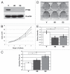
Protein tyrosine kinase 6 (PTK6) is an intracellular tyrosine kinase that is nuclear in epithelial cells of the normal prostate, but cytoplasmic in prostate tumors and in the PC3 prostate tumor cell line. The impact of altered PTK6 intracellular localization in prostate tumor cells has not been extensively explored. Knockdown of endogenous cytoplasmic PTK6 resulted in decreased PC3 cell proliferation and colony formation, suggesting that cytoplasmic PTK6 stimulates oncogenic pathways. In contrast, reintroduction of PTK6 into nuclei of PC3 cells had a negative effect on growth. Enhanced tyrosine phosphorylation of the PTK6 substrate Sam68 was detected in cells expressing nuclear-targeted PTK6. We found that mechanisms regulating nuclear localization of PTK6 are intact in PC3 cells. Transiently overexpressed PTK6 readily enters the nucleus. Ectopic expression of ALT-PTK6, a catalytically inactive splice variant of PTK6, did not affect localization of endogenous PTK6 in PC3 cells. Using leptomycin B, we confirmed that cytoplasmic localization of endogenous PTK6 is not due to Crm-1/exportin-1 mediated nuclear export. In addition, overexpression of the PTK6 nuclear substrate Sam68 is not sufficient to bring PTK6 into the nucleus. While exogenous PTK6 was readily detected in the nucleus when transiently expressed at high levels, low-level expression of inducible wild type PTK6 in stable cell lines resulted in its cytoplasmic retention. Our results suggest that retention of PTK6 in the cytoplasm of prostate cancer cells disrupts its ability to regulate nuclear substrates and leads to aberrant growth. In prostate cancer, restoring PTK6 nuclear localization may have therapeutic advantages.
Figures


Similar articles
-
PTK6 activation at the membrane regulates epithelial-mesenchymal transition in prostate cancer.Cancer Res. 2013 Sep 1;73(17):5426-37. doi: 10.1158/0008-5472.CAN-13-0443. Epub 2013 Jul 15. Cancer Res. 2013. PMID: 23856248 Free PMC article.
-
The alternative splice variant of protein tyrosine kinase 6 negatively regulates growth and enhances PTK6-mediated inhibition of β-catenin.PLoS One. 2011 Mar 30;6(3):e14789. doi: 10.1371/journal.pone.0014789. PLoS One. 2011. PMID: 21479203 Free PMC article.
-
Context-specific protein tyrosine kinase 6 (PTK6) signalling in prostate cancer.Eur J Clin Invest. 2013 Apr;43(4):397-404. doi: 10.1111/eci.12050. Epub 2013 Feb 10. Eur J Clin Invest. 2013. PMID: 23398121 Free PMC article. Review.
-
Protein tyrosine kinase 6 protects cells from anoikis by directly phosphorylating focal adhesion kinase and activating AKT.Oncogene. 2013 Sep 5;32(36):4304-12. doi: 10.1038/onc.2012.427. Epub 2012 Oct 1. Oncogene. 2013. PMID: 23027128 Free PMC article.
-
Brk/PTK6 signaling in normal and cancer cell models.Curr Opin Pharmacol. 2010 Dec;10(6):662-9. doi: 10.1016/j.coph.2010.08.007. Epub 2010 Sep 9. Curr Opin Pharmacol. 2010. PMID: 20832360 Free PMC article. Review.
Cited by
-
PTK6 activation at the membrane regulates epithelial-mesenchymal transition in prostate cancer.Cancer Res. 2013 Sep 1;73(17):5426-37. doi: 10.1158/0008-5472.CAN-13-0443. Epub 2013 Jul 15. Cancer Res. 2013. PMID: 23856248 Free PMC article.
-
BRK targets Dok1 for ubiquitin-mediated proteasomal degradation to promote cell proliferation and migration.PLoS One. 2014 Feb 11;9(2):e87684. doi: 10.1371/journal.pone.0087684. eCollection 2014. PLoS One. 2014. PMID: 24523872 Free PMC article.
-
Downregulated expression of PTK6 is correlated with poor survival in esophageal squamous cell carcinoma.Med Oncol. 2014 Dec;31(12):317. doi: 10.1007/s12032-014-0317-9. Epub 2014 Nov 7. Med Oncol. 2014. PMID: 25377660
-
Targeting protein tyrosine kinase 6 enhances apoptosis of colon cancer cells following DNA damage.Mol Cancer Ther. 2012 Nov;11(11):2311-20. doi: 10.1158/1535-7163.MCT-12-0009. Epub 2012 Sep 18. Mol Cancer Ther. 2012. PMID: 22989419 Free PMC article.
-
HIF-1α-independent hypoxia-induced rapid PTK6 stabilization is associated with increased motility and invasion.Cancer Biol Ther. 2014 Oct;15(10):1350-7. doi: 10.4161/cbt.29822. Epub 2014 Jul 14. Cancer Biol Ther. 2014. PMID: 25019382 Free PMC article.
References
-
- Mitchell PJ, Barker KT, Martindale JE, Kamalati T, Lowe PN, Page MJ, et al. Cloning and characterisation of cDNAs encoding a novel non-receptor tyrosine kinase, brk, expressed in human breast tumours. Oncogene. 1994;9:2383–2390. - PubMed
-
- Siyanova EY, Serfas MS, Mazo IA, Tyner AL. Tyrosine kinase gene expression in the mouse small intestine. Oncogene. 1994;9:2053–2057. - PubMed
-
- Serfas MS, Tyner AL. Brk, Srm, Frk and Src42A form a distinct family of intracellular Src-like tyrosine kinases. Oncol Res. 2003;13:409–419. - PubMed
-
- Barker KT, Jackson LE, Crompton MR. BRK tyrosine kinase expression in a high proportion of human breast carcinomas. Oncogene. 1997;15:799–805. - PubMed
Publication types
MeSH terms
Substances
Grants and funding
LinkOut - more resources
Full Text Sources
Medical
