. 2010 Nov;17(11):1343-51.
doi: 10.1038/nsmb.1911. Epub 2010 Oct 17.

The program for processing newly synthesized histones H3.1 and H4

Affiliations

The program for processing newly synthesized histones H3.1 and H4

Eric I Campos et al. Nat Struct Mol Biol. 2010 Nov.

Abstract

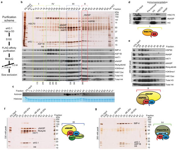
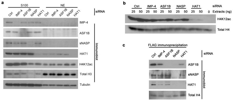
The mechanism by which newly synthesized histones are imported into the nucleus and deposited onto replicating chromatin alongside segregating nucleosomal counterparts is poorly understood, yet this program is expected to bear on the putative epigenetic nature of histone post-translational modifications. To define the events by which naive pre-deposition histones are imported into the nucleus, we biochemically purified and characterized the full gamut of histone H3.1-containing complexes from human cytoplasmic fractions and identified their associated histone post-translational modifications. Through reconstitution assays, biophysical analyses and live cell manipulations, we describe in detail this series of events, namely the assembly of H3-H4 dimers, the acetylation of histones by the HAT1 holoenzyme and the transfer of histones between chaperones that culminates with their karyopherin-mediated nuclear import. We further demonstrate the high degree of conservation for this pathway between higher and lower eukaryotes.


Figures

Figure 1
Figure 1
Subcellular distribution of H3.1-containing complexes. HeLa S3 cells expressing a FLAG-HA tagged histone H3.1 (eH3.1) were fractionated into cytoplasmic (S100), nuclear (NE), and chromatin-bound soluble nuclear pellet (SNP) fractions. (a) Extracts were tandem-affinity purified and resolved by SDS-PAGE for silver staining. (b) Western analysis comparing crude whole cell extracts (WCE), S100, NE, SNP, and total nuclear pellets (TNP) with matching affinity-purified material. (c) Western analysis of endogenous H3.1 co-purifying with exogenous eH3.1 at different cell cycle stages. Arrows mark eH3.1 histones whereas the arrowhead indicates native histone H3.
Figure 2
Figure 2
Purification of eH3.1 from cytosolic extracts. (a) Purification scheme. (b) Anion exchange chromatography (Mono Q) of affinity-purified cytoplasmic eH3.1 protein complexes. Numbers at the bottom indicate the concentration of potassium chloride (mM) at which peak fractions eluted. Cytoplasmic fractions contain four predominant H3.1 complexes (I–IV). (c) Histone acetyltransferase assay using purified histones, the Mono Q fractions, and tritiated acetyl-CoA as substrate. Top panel, autoradiograph. Bottom panel, Coomassie Blue-stained histone substrates. All HAT activity was specific for histone H4. Note that these fraction numbers are displaced by one (odd numbers), relative to those used for the silver stain and western analyses (even numbers). (d) Endogenous HSC70 co-precipitates with endogenous histone H3. Size exclusion chromatography (S200) of the eH3.1 peaks eluting at (e) 380 mM, (f) 330 mM, and (g) 265 mM KCl from the Mono Q column.
Figure 3
Figure 3
sNASP interacts with HAT1-RbAp46 and ASF1B through histone intermediates. (a) FLAG-tagged sNASP and (b) GST-tagged ASF1B were incubated with reciprocal histone chaperones in the presence or absence of H3-H4 and acetyl-CoA. FLAG immunoprecipitates and GST pull-downs, respectively, were analyzed by western blotting.
Figure 4
Figure 4
sNASP homodimerizes but binds H3-H4 heterodimers. (a) Chemical crosslinking of recombinant histones and sNASP or (b) sNASP pre-incubated with H3-H4 at different molar ratios. Crosslinking was performed using wt histones or H3 bearing the indicated point mutations. (c) Sedimentation equilibrium analysis of the complexes between H3-H4 and sNASP. Shown are individual absorbance values at 276 nm recorded at multiple radial positions after equilibrium was reached at 8 krpm (blue), 12 krpm (yellow), and 15 krpm (red). The fitted curves (top graph) are from a single simultaneous fitting of three independent samples at the three speeds. Based on the fixed molecular weights for the postulated species, the data were fitted to the following interaction scheme: (A+A)+B+B⇔A+AB+B⇔(AA)B+B⇔AABB, where A = H3-H4, (AA) is a dimer of A and (A+A) represents self-association, B is the sNASP monomer, AB is the 1:1:1 complex of sNASP:H3:H4, and AABB is its 2:2:2 complex. Bottom graph shows the mean root square deviations.
Figure 5
Figure 5
Importin-4 and ASF1B function downstream of s/tNASP and HAT1 in vivo. (a) Western analysis of H4K12ac levels in cells transfected with siRNA oligos targeting importin-4, ASF1B, NASP (both isoforms), and HAT1. (b) In vitro acetyltransferase activity towards recombinant histones using extracts from cells treated with RNAi against the proteins indicated on top. (c) Immunoprecipitation of histone eH3.1 from cytosolic extracts of RNAi-treated cells.
Figure 6
Figure 6
Conservation of the H3-H4 pre-deposition pathway in S. cerevisiae. (a) TAP-tagged Hif1 was purified from strains wherein different pre-deposition components were deleted as indicated on top, resolved by SDS-PAGE and silver stained (left panel) or analyzed by western (right panel). (b) TAP-tagged Asf1 was purified from strains lacking Hif1, Hat1, or Hat2 and analyzed by western. (c) Histone dependency for Asf1-Hif1 interactions using TAP-tagged versions of wt Asf1 versus the Asf1 mutant V94R.
Figure 7
Figure 7
Model for nuclear import of pre-deposition replication-dependent histones. In humans, H3.1 folding is first assisted by the HSC70 chaperone at the ribosomal exit. H3.1 is transferred to HSP90 that, along with the tNASP co-chaperone, assembles it into H3-H4 units. The sNASP chaperone binds H3.1-H4 heterodimers and presents the H4 carboxyl domain to RbAp46 that recruits HAT1 activity. After acetylation of histone H4, the complex is stabilized and the histones transferred to ASF1B. ASF1B associates with importin-4 and the histones are then transported into the nucleus. In budding yeast, Hif1 associates with the H3-H4 histones and the Hat1-Hat2 holoenzyme. The transfer of acetylated histones to Asf1 is mediated through histones.

References

    1. Eickbush TH, Moudrianakis EN. The histone core complex: an octamer assembled by two sets of protein-protein interactions. Biochemistry. 1978;17:4955–64. - PubMed
    1. Luger K, Mader AW, Richmond RK, Sargent DF, Richmond TJ. Crystal structure of the nucleosome core particle at 2.8 A resolution. Nature. 1997;389:251–60. - PubMed
    1. Campos EI, Reinberg D. Histones: Annotating Chromatin. Annual Review of Genetics. 2009;43:559–599. - PubMed
    1. Hake SB, Allis CD. Histone H3 variants and their potential role in indexing mammalian genomes: the “H3 barcode hypothesis”. Proc Natl Acad Sci U S A. 2006;103:6428–35. - PMC - PubMed
    1. Koessler H, Doenecke D, Albig W. Aberrant expression pattern of replication-dependent histone h3 subtype genes in human tumor cell lines. DNA Cell Biol. 2003;22:233–41. - PubMed

Publication types

MeSH terms