. 2010 Nov;17(11):1337-42.
doi: 10.1038/nsmb.1902. Epub 2010 Oct 17.

Structural basis for cooperative RNA binding and export complex assembly by HIV Rev

Affiliations

Structural basis for cooperative RNA binding and export complex assembly by HIV Rev

Matthew D Daugherty et al. Nat Struct Mol Biol. 2010 Nov.

Abstract

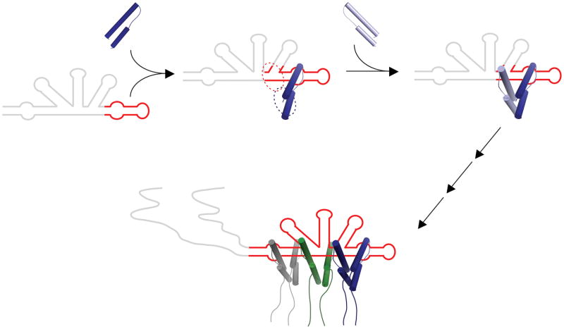
HIV replication requires nuclear export of unspliced viral RNAs to translate structural proteins and package genomic RNA. Export is mediated by cooperative binding of the Rev protein to the Rev response element (RRE) RNA, to form a highly specific oligomeric ribonucleoprotein (RNP) that binds to the Crm1 host export factor. To understand how protein oligomerization generates cooperativity and specificity for RRE binding, we solved the crystal structure of a Rev dimer at 2.5-Å resolution. The dimer arrangement organizes arginine-rich helices at the ends of a V-shaped assembly to bind adjacent RNA sites and structurally couple dimerization and RNA recognition. A second protein-protein interface arranges higher-order Rev oligomers to act as an adaptor to the host export machinery, with viral RNA bound to one face and Crm1 to another, the oligomers thereby using small, interconnected modules to physically arrange the RNP for efficient export.


Conflict of interest statement

The authors declare no conflict of interest.

Figures

Figure 1
Figure 1
Rev domain structure and protein dimerization. (a) Domain structure of the Rev protein, including the disordered C-terminal region containing the nuclear export sequence (NES). Below is a consensus alignment of the first 70 residues of Rev from reference subtypes (Los Alamos HIV sequence database (http://www.hiv.lanl.gov/)) represented using the ClustalW program. The top sequence is the one used for crystallization, with oligomerization mutants L12S and L60R indicated by daggers, and above is the predicted secondary structure. Regions predicted to be helical are indicated by wavy lines and coiled regions by dashed lines. Highlighted are residues shown in the Rev dimer structure to be important for Rev monomer stability and folding (blue), Rev dimer formation (yellow), or both (green). Residues in red are the highly conserved residues of the ARM. (b) Representative gel shift assay data using purified Rev70-Dimer at the concentrations indicated and radiolabeled IIB42 RNA. The species indicated are: F, Free RNA; M, Rev monomer; D, Rev dimer. The dissociation constant (KD = 14 ± 6.7 nM) and Hill coefficient (η = 1.1 ± 0.027) were calculated using the equation: FractionBound = [Rev]η/(KDη+[Rev]η). Data are presented as the mean ± s.d. from three replicates. (c) Measured multi-angle light scattering (MALS) (dashed lines, left axis) and calculated molar masses (solid line, right axis) determined from the major peak from size exclusion chromatography (SEC) of 250 μM Rev70-Dimer protein in crystallization buffer containing 0.5 M SO4. The measured mass of 17 kDa corresponds to two Rev monomers (8.5 kDa each).
Figure 2
Figure 2
Overall structure of the Rev dimer and monomer. (a) Two views of a surface representation of the Rev dimer, with the two monomers shown as ribbons (dark and light blue) and the crossing angle indicated. (b) Each of the four Rev monomers has a folded core from residues 9–63 as shown, with structural and functional regions and amino acid numberings indicated. All monomer structures are highly similar, with a root mean squared deviation (r.m.s.d.) of 0.5–1 Å for all pairwise alignments of backbone atoms from residues 9–63, and a backbone r.m.s.d of ≤1.0 Å when aligned to the recent Fab-bound Rev structure. Amino acid conservation among 1201 HIV-1 isolates in the Los Alamos HIV sequence database is indicated on the ribbon diagram, ranging from lowest conservation (26%, green) to highest (100%, red). Hydrophobic and polar residues that stabilize the monomer structure are shown as spheres and sticks and are colored by sequence conservation. (c) Stereo view of a σA-weighted 2Fobs–Fcalc map contoured at 2.0σ of the hydrogen bonding network stabilizing the Rev monomer structure. Distances between hydrogen-bonding donor and acceptor atoms are indicated (Å).
Figure 3
Figure 3
The Rev dimer interface mediates cooperative RNA recognition. (a) Cutaway of the dimer interface showing the surface of one monomer (blue), and the predicted solvent accessible surface area buried upon dimerization (orange) with the second monomer (light blue ribbon). (b) The five critical hydrophobic residues that comprise the dimer interface, shown as sticks in one monomer projecting onto the surface of the second. (c) Three hydrophobic residues mediate symmetric interactions at the dimerization interface. Individual monomers are colored in light and dark blue. (d) Residues that form the core of the monomer structure (Leu22 and Ile55) are buried further upon dimerization. Nonpolar surface area of these residues is partially buried in the monomer (blue) and additionally buried in the dimer (orange). (e) Two views of the Rev dimer (blue) modeled with the 34 nt stem IIB (labeled IIB34; red) from the NMR structure of an ARM peptide–RNA complex. The peptide helix was aligned with ARM1 (r.m.s.d = 1.0 Å), and four additional base pairs of idealized A-form RNA were appended to the end of the RNA (red). The 42 nt RNA that binds the Rev dimer cooperatively (Fig. 1) contains bulged nucleotides that are missing from this model, but the trajectory and length of the helix should be similar. The positions of Asn40 are shown for each monomer (yellow), showing how the ‘inner face’ of ARM1 positions Asn40 to contact stem IIB while the ‘outer face’ of ARM2 (without Asn40) is positioned to contact the adjacent RNA site.
Figure 4
Figure 4
Arrangement of the Rev oligomer and model of the Rev–RRE RNP and Crm1 interaction. (a) Comparison of the higher-order oligomerization interface seen between asymmetric units in the crystal (blue and green cartoon) and a recent structure of Rev with a monoclonal Fab bound to the dimer interface (grey cylinders) (backbone r.m.s.d = 1.9 A). (b) A Rev hexamer generated using the arrangement of three Rev dimers in the crystal (blue, green and light blue cartoons). (c) Two views of a jellyfish model of the Rev hexamer bound to RNA and its interaction with Crm1. RNA was docked onto one dimer of the Rev hexamer as in Fig. 2e, and extended an additional 9 base pairs of A-form helix to contact the second dimer (seen in the right panel, green dimer) based on previous studies with model RNAs. An additional fragment of RNA was docked in the same manner as in Fig. 2e to the third dimer (light blue) to model complete binding of the Rev hexamer to the RRE (see Methods). The disordered C-termini containing the NES (residues 73–83; orange) were modeled as random coil extensions and are seen to project away from the RNA-binding ARMs. The NES sequences were fit into the NES binding site (cyan) of Crm1 based on the structure of a Crm1–RanGTP complex (dark and light grey surfaces, respectively) with Snurportin1 bound at the NES site. Two Crm1 complexes are shown for scale and suggest that no more than two are likely to be accommodated in this arrangement.
Figure 5
Figure 5
Model for oligomerization-mediated cooperative assembly. Binding of Rev (top left, blue cartoon) to the RRE (grey) is initiated at the stem IIB site (red). The resulting complex (top middle) exposes the dimerization interface (blue dashed oval) for binding by a second Rev monomer. Only when a second Rev-binding RNA site (red dashed oval) is properly oriented relative to the stem IIB site will the second Rev monomer be able to cooperatively assemble into the RNP using this composite protein–protein and protein–RNA interaction surface. Further oligomerization and protein–RNA interactions will likewise facilitate cooperative assembly of the complete hexameric Rev–RRE RNP (bottom). At this point, the complex is organized for nuclear export, with the NESs of the Rev subunits projecting downward (as in Fig. 4c) for interaction with Crm1 and the viral RNA bound to opposite face of the Rev oligomer.

References

    1. Cullen BR. Nuclear mRNA export: insights from virology. Trends Biochem Sci. 2003;28:419–24. - PubMed
    1. Pollard VW, Malim MH. The HIV-1 Rev Protein. Annu Rev Microbiol. 1998;52:491–532. - PubMed
    1. Fornerod M, Ohno M, Yoshida M, Mattaj IW. CRM1 is an export receptor for leucine-rich nuclear export signals. Cell. 1997;90:1051–1060. - PubMed
    1. Malim MH, Cullen BR. HIV-1 structural gene expression requires the binding of multiple Rev monomers to the viral RRE: implications for HIV-1 latency. Cell. 1991;65:241–48. - PubMed
    1. Mann DA, et al. A molecular rheostat: Co-operative rev binding to stem I of the Rev-response element modulates human immunodeficiency virus type-1 late gene expression. J Mol Biol. 1994;241:193–207. - PubMed

Publication types

MeSH terms

Substances

Associated data