. 2011:2011:739093.
doi: 10.1155/2011/739093. Epub 2010 Sep 26.

Ocimum gratissimum Aqueous Extract Induces Apoptotic Signalling in Lung Adenocarcinoma Cell A549

Affiliations

Ocimum gratissimum Aqueous Extract Induces Apoptotic Signalling in Lung Adenocarcinoma Cell A549

Han-Min Chen et al. Evid Based Complement Alternat Med. 2011.

Abstract

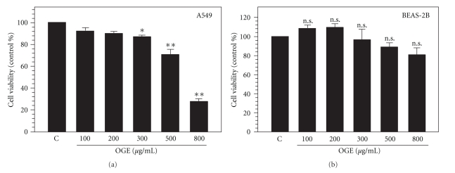
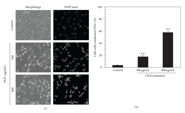
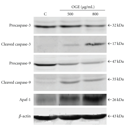
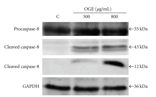
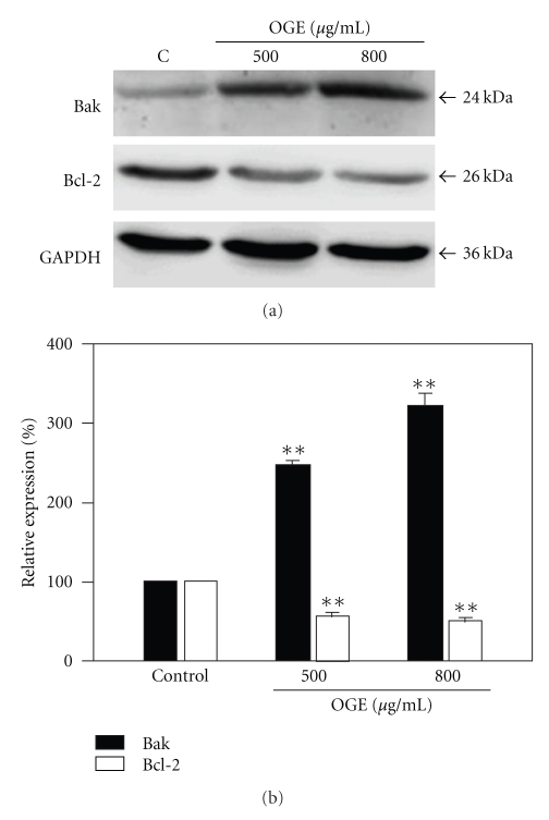
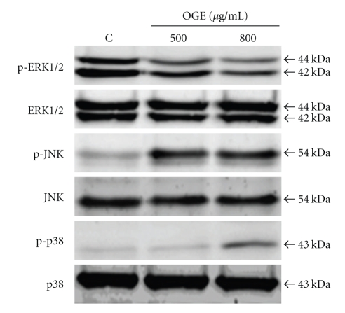
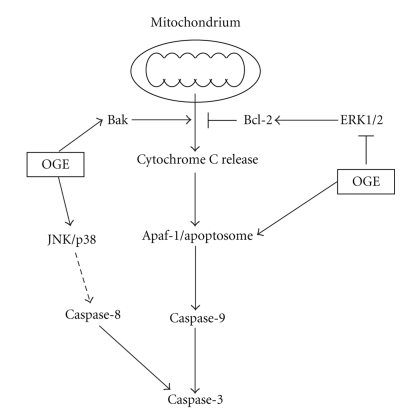
Ocimum gratissimum (OG) is widely used as a traditional herb for its antibacterial activity in Taiwan. Recently, antitumor effect of OG on breast cancer cell is also reported; however, the effects of OG on human pulmonary adenocarcinoma cell A549 remain unclear. Therefore, we aimed to investigate whether aqueous OG extract (OGE) affects viability of A549 cells and the signals induced by OGE in A549 cells. Cell viability assays revealed that OGE significantly and dose-dependently decreased the viability of A549 cell but not that of BEAS-2B cell. Morphological examination and DAPI staining indicated that OGE induced cell shrinkage and DNA condensation for A549 cells. Further investigation showed that OGE enhanced activation of caspase-3, caspase-9 and caspase-8 and increased protein level of Apaf-1 and Bak, but diminished the level of Bcl-2. Additionally, OGE inhibited the phosphorylation of extracellular signal-regulated kinase (ERK) yet enhanced the phosphorylation of c-Jun N-terminal kinase (JNK) and p38 MAP kinase (p38). In conclusion, our findings indicate that OGE suppressed the cell viability of A549 cells, which may result from the activation of apoptotic signaling and the inhibition of anti-apoptotic signaling, suggesting that OGE might be beneficial to lung carcinoma treatment.


Figures

Figure 1
Figure 1
OGE diminished cell viability of A549 cell. The cell viability of A549 cells (a) and BEAS-2B cells (b) treated with serial concentrations of OGE (10, 50, 100, 200 and 300 μg/ml) for 48 h was determined. Data were expressed as mean ±  SEM for 3 independent experiments. *P < .05 and, **P < .01 as comparing to control (c). n.s., no statistical significance.
Figure 2
Figure 2
OGE altered cell morphology and induced DNA condensation of A549 cell. A549 cells were treated with 0, 500 and 800 μg/ml OGE for 48 h and then stained with DAPI. (a) The cell morphology and the DNA condensation was photographed by fluorescence microscopy (200x). The cells presented DNA condensation were indicated by arrow. (b) The incidence of DNA condensation was determined for the A549 cells with different treatments. Data were expressed as mean ±  SEM for 3 independent experiments.
Figure 3
Figure 3
OGE induced activation of intrinsic/mitochondrial apoptotic pathway. A549 cells were treated with 0, 500 and 800 μg/ml for 48 hr, and then were lyzed for the determination of protein levels of caspase-3, cleaved caspase-3, caspase-9, cleaved caspase-9 and Apaf-1 by immunoblotting. β-actin was used as control. The apparent molecular weights for detected proteins were indicated.
Figure 4
Figure 4
OGE induced activation of caspase-8. A549 cells were treated with 0, 500 and 800 μg/ml for 48 hr, and then were lyzed for the determination of protein levels of caspase-8 and cleaved caspase-8 by immunoblotting. β  actin was used as control. The apparent molecular weights for detected proteins were indicated.
Figure 5
Figure 5
OGE enhanced protein level of Bak and diminished protein level of Bcl-2. A549 cells were treated with 0, 500, and 800 μg/ml for 48 hr, and then were lyzed for the determination of protein levels by immunoblotting. (a) The expression levels of Bak and Bcl-2 were determined. GAPDH was used as control. (b) The expression levels of Bak and Bcl-2 were quantitatively expressed after being standardized to GAPDH. Data are expressed as mean ±  SEM for 3 independent experiments for each concentration point. **P < .01 as compared with control.
Figure 6
Figure 6
OGE inhibited phosphorylation of Erk but enhanced phosphorylation of JNK and p38. A549 cells were treated with 0, 500 and 800 μg/ml for 48 hr, and then were lyzed for the determination of protein levels by immunoblotting. The levels of p-ERK1/2, ERK1/2, p-JNK, JNK, p-p38, and p38 were presented.
Figure 7
Figure 7
Proposed model for the antitumoral activity of Ocimum gratissimum extracts on the human lung adenocarcinoma A549. Our data demonstrated that Ocimum gratissimum extract induces activation of caspase-3, caspase-9, and caspase-8, which may attribute to the increase of Apaf-1 and Bak, the decrease of antiapoptotic Bcl-2, as well as the inhibition of ERK1/2 survival signaling and the enhancement of JNK and p38 stress signal cascades.

Similar articles

Cited by

References

    1. Jemal A, Siegel R, Ward E, et al. Cancer statistics, 2006. Ca: A Cancer Journal for Clinicians. 2006;56(2):106–130. - PubMed
    1. Parkin DM. Global cancer statistics in the year 2000. Lancet Oncology. 2001;2(9):533–543. - PubMed
    1. Zhu W, Wang X-M, Zhang L, Li X-Y, Wang B-X. Pharmacokinetic of Rhein in healthy male volunteers following oral and retention enema administration of rhubarb extract: a single dose study. American Journal of Chinese Medicine. 2005;33(6):839–850. - PubMed
    1. Willett WC. Diet and health: what should we eat? Science. 1994;264(5158):532–537. - PubMed
    1. Fontham ETH. Protective dietary factors and lung cancer. International Journal of Epidemiology. 1990;19(1):S32–S42. - PubMed