Beneficial effect of the traditional chinese drug shu-xue-tong on recovery of spinal cord injury in the rat
- PMID: 20953395
- PMCID: PMC2952331
- DOI: 10.1155/2011/862197
Beneficial effect of the traditional chinese drug shu-xue-tong on recovery of spinal cord injury in the rat
Abstract
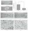
Shu-Xue-Tong (SXT) is a traditional Chinese drug widely used to ameliorate stagnation of blood flow, such as brain or myocardial infarction. Whether SXT may have therapeutic value for spinal cord injury (SCI), during which ischemia plays an important role in its pathology, remains to be elucidated. We hypothesized that SXT may promote SCI healing by improving spinal cord blood flow (SCBF), and a study was thus designed to explore this possibility. Twenty-five male Sprague-Dawley rats were used. SCI was induced by compression, and SXT was administrated 24 h postinjury for 14 successive days. The effects of SXT were assessed by means of laser-Doppler flowmetry, motor functional analysis (open-field walking and footprint analysis), and histological analysis (hematoxylin-eosin and thionin staining and NeuN immunohistochemistry). SXT significantly promoted SCBF of the contused spinal cord and enhanced the recovery of motor function. Histological analysis indicated that the lesion size was reduced, the pathological changes were ameliorated, and more neurons were preserved. Based on these results we conclude that SXT can effectively improve SCI.
Figures


Similar articles
-
Protective effects of Batroxobin on spinal cord injury in rats.Neurosci Bull. 2013 Aug;29(4):501-8. doi: 10.1007/s12264-013-1354-7. Epub 2013 Jul 13. Neurosci Bull. 2013. PMID: 23852558 Free PMC article.
-
Low-energy extracorporeal shock wave therapy promotes vascular endothelial growth factor expression and improves locomotor recovery after spinal cord injury.J Neurosurg. 2014 Dec;121(6):1514-25. doi: 10.3171/2014.8.JNS132562. Epub 2014 Oct 3. J Neurosurg. 2014. PMID: 25280090
-
The Effect of Shu Xue Tong Treatment on Random Skin Flap Survival via the VEGF-Notch/Dll4 Signaling Pathway.J Invest Surg. 2020 Aug;33(7):615-620. doi: 10.1080/08941939.2018.1551948. Epub 2019 Jan 15. J Invest Surg. 2020. PMID: 30644800
-
Neurological recovery is impaired by concurrent but not by asymptomatic pre-existing spinal cord compression after traumatic spinal cord injury.Spine (Phila Pa 1976). 2012 Aug 1;37(17):1448-55. doi: 10.1097/BRS.0b013e31824ffda5. Spine (Phila Pa 1976). 2012. PMID: 22414995
-
Mash-1 modified neural stem cells transplantation promotes neural stem cells differentiation into neurons to further improve locomotor functional recovery in spinal cord injury rats.Gene. 2021 May 20;781:145528. doi: 10.1016/j.gene.2021.145528. Epub 2021 Feb 22. Gene. 2021. PMID: 33631250
Cited by
-
Spinal cord contusion.Neural Regen Res. 2014 Apr 15;9(8):789-94. doi: 10.4103/1673-5374.131591. Neural Regen Res. 2014. PMID: 25206890 Free PMC article. Review.
-
Protective effects of Batroxobin on spinal cord injury in rats.Neurosci Bull. 2013 Aug;29(4):501-8. doi: 10.1007/s12264-013-1354-7. Epub 2013 Jul 13. Neurosci Bull. 2013. PMID: 23852558 Free PMC article.
-
A Parallel-Arm Randomized Controlled Trial to Assess the Effects of a Far-Infrared-Emitting Collar on Neck Disorder.Materials (Basel). 2015 Sep 1;8(9):5862-5876. doi: 10.3390/ma8095279. Materials (Basel). 2015. PMID: 28793539 Free PMC article.
-
Different TLR4 expression and microglia/macrophage activation induced by hemorrhage in the rat spinal cord after compressive injury.J Neuroinflammation. 2013 Sep 10;10:112. doi: 10.1186/1742-2094-10-112. J Neuroinflammation. 2013. PMID: 24015844 Free PMC article.
-
Early Blockade of TLRs MyD88-Dependent Pathway May Reduce Secondary Spinal Cord Injury in the Rats.Evid Based Complement Alternat Med. 2012;2012:591298. doi: 10.1155/2012/591298. Epub 2012 May 22. Evid Based Complement Alternat Med. 2012. PMID: 22675384 Free PMC article.
References
-
- McDonald JW, Sadowsky C. Spinal-cord injury. Lancet. 2002;359(9304):417–425. - PubMed
-
- Schwab JM, Brechtel K, Mueller C-A, et al. Experimental strategies to promote spinal cord regeneration—an integrative perspective. Progress in Neurobiology. 2006;78(2):91–116. - PubMed
-
- Profyris C, Cheema SS, Zang D, Azari MF, Boyle K, Petratos S. Degenerative and regenerative mechanisms governing spinal cord injury. Neurobiology of Disease. 2004;15(3):415–436. - PubMed
-
- Carlson GD, Gorden C. Current developments in spinal cord injury research. Spine Journal. 2002;2(2):116–128. - PubMed
-
- Nashmi R, Fehlings MG. Mechanisms of axonal dysfunction after spinal cord injury: with an emphasis on the role of voltage-gated potassium channels. Brain Research Reviews. 2001;38(1-2):165–191. - PubMed
LinkOut - more resources
Full Text Sources

