

In vivo imaging biomarkers in mouse models of Alzheimer's disease: are we lost in translation or breaking through?
- PMID: 20953404
- PMCID: PMC2952791
- DOI: 10.4061/2010/604853
In vivo imaging biomarkers in mouse models of Alzheimer's disease: are we lost in translation or breaking through?
Abstract
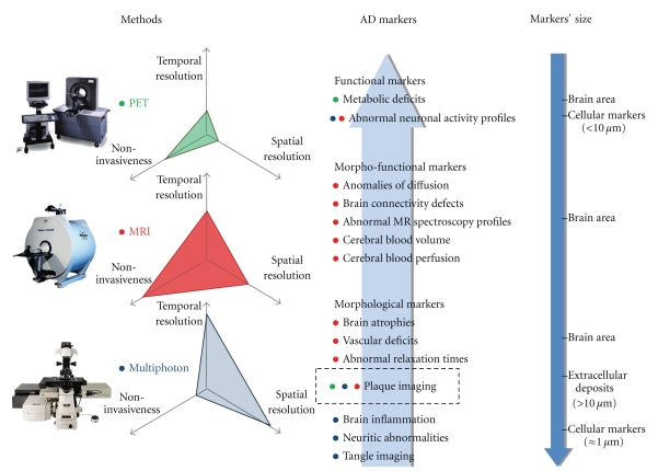
Identification of biomarkers of Alzheimer's Disease (AD) is a critical priority to efficiently diagnose the patients, to stage the progression of neurodegeneration in living subjects, and to assess the effects of disease-modifier treatments. This paper addresses the development and usefulness of preclinical neuroimaging biomarkers of AD. It is today possible to image in vivo the brain of small rodents at high resolution and to detect the occurrence of macroscopic/microscopic lesions in these species, as well as of functional alterations reminiscent of AD pathology. We will outline three different types of imaging biomarkers that can be used in AD mouse models: biomarkers with clear translational potential, biomarkers that can serve as in vivo readouts (in particular in the context of drug discovery) exclusively for preclinical research, and finally biomarkers that constitute new tools for fundamental research on AD physiopathogeny.
Figures
References
-
- Sarazin M, Berr C, De Rotrou J, et al. Amnestic syndrome of the medial temporal type identifies prodromal AD: a longitudinal study. Neurology. 2007;69(19):1859–1867. - PubMed
-
- Gearing M, Mirra SS, Hedreen JC, Sumi SM, Hansen LA, Heyman A. The Consortium to Establish a Registry for Alzheimer’s Disease (CERAD)—part X: neuropathology confirmation of the clinical diagnosis of Alzheimer’s disease. Neurology. 1995;45(3):461–466. - PubMed
-
- Edison P, Archer HA, Gerhard A, et al. Microglia, amyloid, and cognition in Alzheimer’s disease: an [11C](R)PK11195-PET and [11C]PIB-PET study. Neurobiology of Disease. 2008;32(3):412–419. - PubMed
-
- Scheltens P, Korf ESC. Contribution of neuroimaging in the diagnosis of Alzheimer’s disease and other dementias. Current Opinion in Neurology. 2000;13(4):391–396. - PubMed
LinkOut - more resources
Full Text Sources
