

Systemic lupus erythematosus monocytes are less responsive to interleukin-10 in the presence of immune complexes
- PMID: 20954190
- PMCID: PMC3014998
- DOI: 10.1002/art.30083
Systemic lupus erythematosus monocytes are less responsive to interleukin-10 in the presence of immune complexes
Abstract
Objective: Systemic lupus erythematosus (SLE) is a systemic inflammatory disease characterized by autoantibody production and immune complex deposition. The level of interleukin-10 (IL-10), predominantly an antiinflammatory cytokine, is paradoxically elevated in patients with SLE. The aim of this study was to examine the hypothesis that the antiinflammatory function of IL-10 is impaired in monocytes from patients with SLE with long-term exposure to immune complexes.
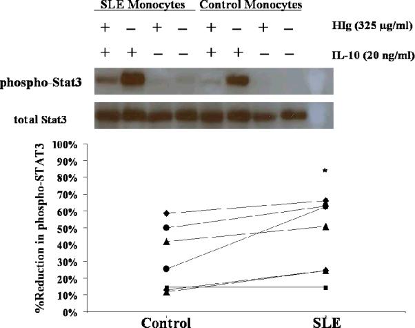
Methods: CD14+ monocytes were isolated from healthy donors and patients with SLE. Cultured CD14+ cells were treated with heat-aggregated human IgG (325 μg/ml) in the presence or absence of IL-10 (20 ng/ml). To study gene expression, RNA was extracted 3 hours after treatment. To study cytokine production, supernatants were harvested after 8 hours. To study IL-10 signaling, cell lysates were obtained from CD14+ cells treated with human IgG (325 μg/ml) for 1 hour followed by IL-10 (20 ng/ml) treatment for 10 minutes. Western blot analysis was used to assess STAT-3 phosphorylation. All experiments were performed in pairs.
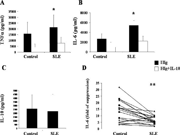
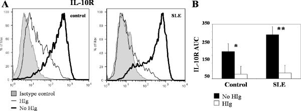
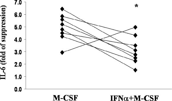
Results: When stimulated with human IgG, SLE monocytes produced more tumor necrosis factor α (TNFα) and IL-6 than did control cells. The suppressive effect of IL-10 on human IgG-induced TNFα and IL-6 production was lower in SLE monocytes compared with control monocytes, although IL-10 receptor expression was similar in SLE and control monocytes. Human IgG suppressed IL-10 receptor expression and altered IL-10 signaling in control monocytes. Like SLE monocytes, interferon-α (IFNα)-primed control monocytes stimulated with human IgG were also less responsive to IL-10.
Conclusion: Human IgG and IFNα modulate IL-10 function. In SLE monocytes, which are considered to be IFNα primed and persistently exposed to immune complexes, responses to IL-10 are abnormal, limiting the antiinflammatory effect of this cytokine.
Copyright © 2011 by the American College of Rheumatology.
Figures
References
-
- Moore KW, de Waal Malefyt R, Coffman RL, O'Garra A. Interleukin-10 and the interleukin-10 receptor. Annual Review of Immunology. 2001;19:683–765. - PubMed
-
- Kasama T, Strieter RM, Lukacs NW, Burdick MD, Kunkel SL. Regulation of neutrophil-derived chemokine expression by IL-10. J Immunol. 1994;152(7):3559–69. - PubMed
-
- Ding L, Linsley PS, Huang LY, Germain RN, Shevach EM. IL-10 inhibits macrophage costimulatory activity by selectively inhibiting the up-regulation of B7 expression. J Immunol. 1993;151(3):1224–34. - PubMed
-
- Houssiau FA, Lefebvre C, Vanden Berghe M, Lambert M, Devogelaer JP, Renauld JC. Serum interleukin 10 titers in systemic lupus erythematosus reflect disease activity. Lupus. 1995;4(5):393–5. - PubMed
-
- Lazarus M, Hajeer AH, Turner D, Sinnott P, Worthington J, Ollier WE. Genetic variation in the interleukin 10 gene promoter and systemic lupus erythematosus. J Rheumatol. 1997;24(12):2314–7. - PubMed
Publication types
MeSH terms
Substances
Grants and funding
LinkOut - more resources
Full Text Sources
Medical
Research Materials
Miscellaneous
