

Structural neuroimaging in schizophrenia: from methods to insights to treatments
- PMID: 20954428
- PMCID: PMC3181976
- DOI: 10.31887/DCNS.2010.12.3/mshenton
Structural neuroimaging in schizophrenia: from methods to insights to treatments
Abstract
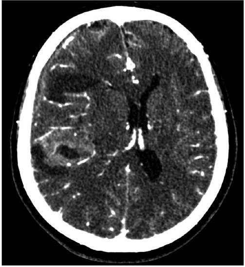
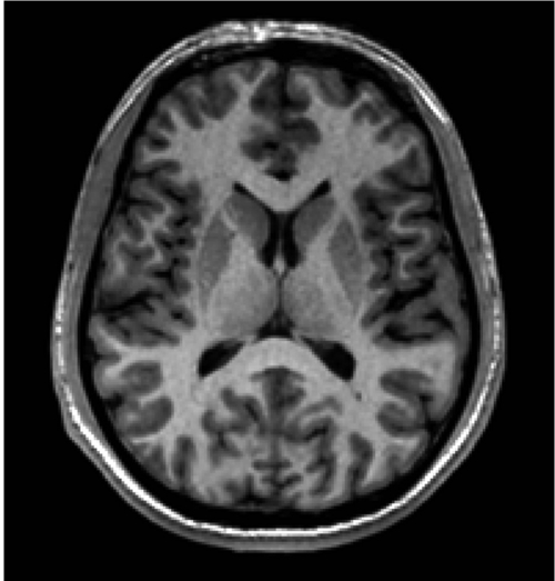
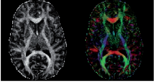
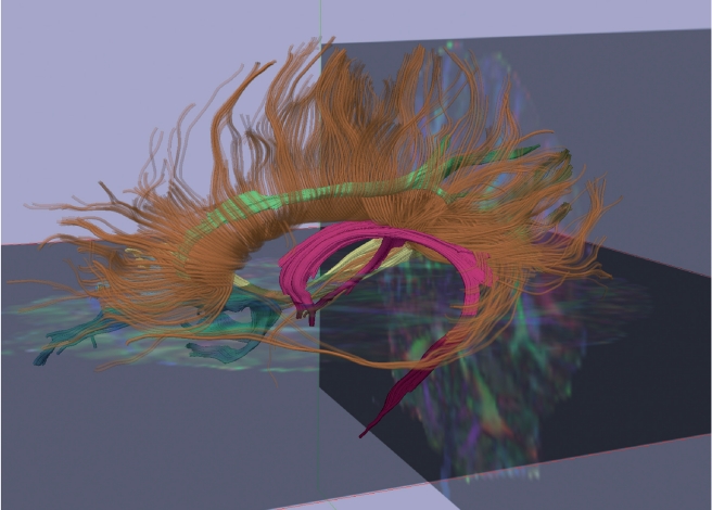
Historically, Kraepelin speculated that dementia praecox resulted from damage to the cerebral cortex, most notably the frontal and temporal cortices. It is only recently, however, that tools have been available to test this hypothesis. Now, more than a century later, we know that schizophrenia is a brain disorder. This knowledge comes from critical advances in imaging technology--including computerized axial tomography, magnetic resonance imaging, and diffusion imaging--all of which provide an unprecedented view of neuroanatomical structures, in vivo. Here, we review evidence for structural neuroimaging abnormalities, beginning with evidence for focal brain abnormalities, primarily in gray matter, and proceeding to the quest to identify abnormalities in brain systems and circuits by focusing on damage to white matter connections in the brain. We then review future prospects that need to be explored and pursued in order to translate our current knowledge into an understanding of the neurobiology of schizophrenia, which can then be translated into novel treatments.
Desde un punto de vista histórico Kraepelin formuló la hipótesis que la demencia precoz se producía por un daño en la corteza cerebral, más específicamente en las cortezas frontal y temporal. Sin embargo, sólo recientemente se ha podido disponer de herramientas para evaluar esta hipótesis. Desde hace más de un siglo que ya se sabe que la esquizofrenia es un trastorno cerebral. Este conocimiento procede de importantes avances en la tecnología de imágenes -que incluyen la tomografía axial computarizada, las imágenes de resonancia magnética y las imágenes de difusión- todos los cuales aportan una visión, in vivo, sin precedentes de las estructuras neuroanatómicas. En este artículo se revisan las evidencias de las anormalidades estructurales de las neuroimágenes, comenzando con evidencias de alteraciones cerebrales focales, principalmente en la sustancia gris y continuando con las investigaciones para identificar anormalidades en los sistemas y circuitos cerebrales, focalizándose en el daño en las conexiones con la sustancia blanca del cerebro. Luego se revisan futuras líneas de investigación que requieren ser exploradas y desarrolladas para traducir el conocimiento actual en una comprensión de la neurobiología de la esquizofrenia, que puede traducirse en nuevos tratamientos.
Historiquement, Kraepelin a émis l'hypothèse que la démence précoce résultait d'une lésion du cortex cérébral, plus particulièrement du cortex frontal et du cortex temporal. Ce n'est cependant que récemment seulement que des outils ont permis de tester cette hypothèse. Maintenant, plus d'un siècle plus tard, nous savons que la schizophrénie est un trouble cérébral grâce à des avancées décisives en imagerie, comprenant la tomographie axiale numérisée, l'imagerie par résonance magnétique et l'imagerie de diffusion, toutes fournissant un aperçu sans précédent des structures neuroanatomiques in vivo. Nous présentons ici les données scientifiques en faveur de l'existence d'anomalies identifiées par la neuro-imagerie structurelle, en commençant par les anomalies cérébrales focales, tout d'abord dans la substance grise et en poursuivant par l'identification des anomalies des circuits et des systèmes cérébraux en s'attachant en particulier aux lésions des connexions de la substance blanche cérébrale. Puis nous abordons les futures perspectives à explorer afin de transformer notre connaissance actuelle en une compréhension de la neurobiologie de la schizophrénie, afin de développer par la suite de nouveaux traitements.
Figures
References
-
- Kraepelin E. Dementia Praecox. In: Barclay BBE, ed, tran . 1919-1971 New York, NY: Churchill Livingston Inc;
-
- Bleuler E. Dementia Praecox or The Group of Schizophrenias. New York, NY: International Universities Press . 1911-1950
-
- Benes F. Is there a neuroanatomical basis for schizophrenia? An old question revisited. Neuroscientist. 1995;1:104–115.
-
- Bogerts B. Neuropathology of schizophrenias. Fortschr Neurol Psychiatr. 1984;52:428–437. - PubMed
Publication types
MeSH terms
Grants and funding
LinkOut - more resources
Full Text Sources
Medical