

Exploring cellular contact guidance using gradient nanogratings
- PMID: 20954734
- PMCID: PMC3061972
- DOI: 10.1021/bm100883m
Exploring cellular contact guidance using gradient nanogratings
Abstract
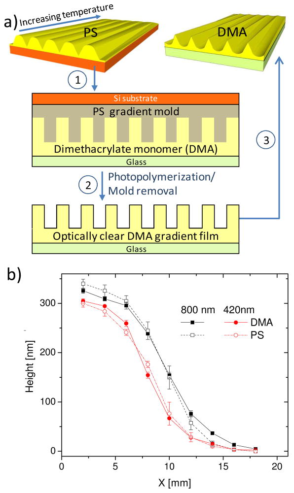
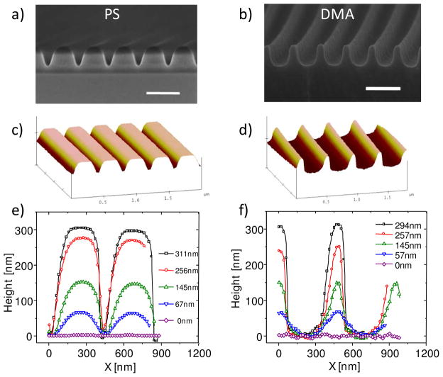
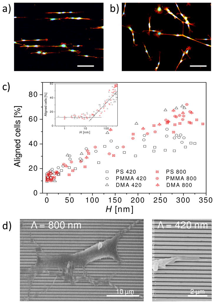
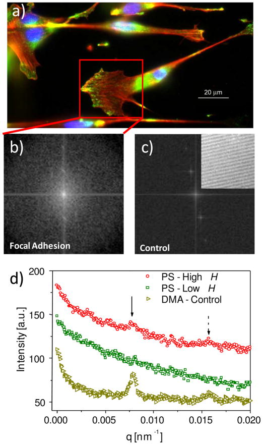
Nanoscale surface features that mimic extracellular matrix are critical environmental cues for cell contact guidance and are vital in advanced medical devices in order to manipulate cell behaviors. Among them, nanogratings (line-and-space gratings) are common platforms to study geometric effects on cell contact guidance, especially cell alignment, but generally are one pattern height per platform. In this study, we developed a strategy to fabricate controlled substrates with a wide range of pattern shapes and surface chemistries and to separate surface chemistry and topography effects. As a demonstration of this strategy, six nanograting platforms on three materials were fabricated and applied to examine and differentiate the effects of surface topography and surface chemistry on cell contact guidance of murine preosteoblasts. All of the six platforms contained the same gradient in pattern height (0 to ≈350 nm). They were prepared using nanoimprint lithography and annealing for thermoplastic materials (low molecular weight polystyrene (PS) and polymethylmethacrylate (PMMA)) and photoimprint for a thermoset material (a cross-linked dimethacrylate (DMA)). Each material contains two platforms that are only different in line-and-space pitch (420 or 800 nm). The DMA nanogratings had a reverse line-and-space profile to those of the PS and PMMA nanogratings. Using these platforms, a full range of cell alignment, from randomly orientated to completely parallel to the grating direction was achieved. Results from focal adhesion assays and scanning electronic microscopy indicated a change in cell-substrate contact from a noncomposite state (full contact) to a composite state (partial contact between cell and substrate) as pattern height increased. These gradient platforms allowed for the separation of surface chemistry and surface topography to provide insight into the mechanisms responsible for cell contact guidance on nanopatterned surfaces.
Figures
Similar articles
-
Superhydrophobic Surfaces on Phase-separated Nanostructures of Polystyrene/Polymethyl Methacrylate Films Fabricated by the Double-spray Technique.J Oleo Sci. 2018 Sep 1;67(9):1101-1105. doi: 10.5650/jos.ess18061. Epub 2018 Aug 14. J Oleo Sci. 2018. PMID: 30111680
-
Nanotopographical guidance of C6 glioma cell alignment and oriented growth.Biomaterials. 2004 Aug;25(18):4215-23. doi: 10.1016/j.biomaterials.2003.11.020. Biomaterials. 2004. PMID: 15046911
-
Nanoscale rings fabricated using self-assembled triblock terpolymer templates.ACS Nano. 2008 Oct 28;2(10):2007-14. doi: 10.1021/nn8002345. ACS Nano. 2008. PMID: 19206445
-
Fabrication of hierarchical micro-nanotopographies for cell attachment studies.Nanotechnology. 2013 Jun 28;24(25):255305. doi: 10.1088/0957-4484/24/25/255305. Epub 2013 May 31. Nanotechnology. 2013. PMID: 23727615
-
Nanopatterned polymer brushes: conformation, fabrication and applications.Nanoscale. 2016 Jan 14;8(2):680-700. doi: 10.1039/c5nr07107k. Nanoscale. 2016. PMID: 26648412 Review.
Cited by
-
Deterministic Integration of Biological and Soft Materials onto 3D Microscale Cellular Frameworks.Adv Biosyst. 2017 Sep;1(9):1700068. doi: 10.1002/adbi.201700068. Epub 2017 Jul 31. Adv Biosyst. 2017. PMID: 29552634 Free PMC article.
-
High-Performance Dental Adhesives Containing an Ether-Based Monomer.J Dent Res. 2020 Feb;99(2):189-195. doi: 10.1177/0022034519895269. Epub 2019 Dec 20. J Dent Res. 2020. PMID: 31861961 Free PMC article.
-
Dynamic Manipulation of Cell Membrane Curvature by Light-Driven Reshaping of Azopolymer.Nano Lett. 2020 Jan 8;20(1):577-584. doi: 10.1021/acs.nanolett.9b04307. Epub 2019 Dec 19. Nano Lett. 2020. PMID: 31846332 Free PMC article.
-
Nanoscale Surface Topography Reduces Focal Adhesions and Cell Stiffness by Enhancing Integrin Endocytosis.Nano Lett. 2021 Oct 13;21(19):8518-8526. doi: 10.1021/acs.nanolett.1c01934. Epub 2021 Aug 4. Nano Lett. 2021. PMID: 34346220 Free PMC article.
-
Engineered 3D Polymer and Hydrogel Microenvironments for Cell Culture Applications.Bioengineering (Basel). 2019 Dec 13;6(4):113. doi: 10.3390/bioengineering6040113. Bioengineering (Basel). 2019. PMID: 31847117 Free PMC article. Review.
References
-
- Stevens MM, George JH. Science. 2005;310(5751):1135–1138. - PubMed
-
- Cukierman E, Pankov R, Stevens DR, Yamada KM. Science. 2001;294(5547):1708–1712. - PubMed
-
- Langer R, Vacanti JP. Science. 1993;260(5110):920–926. - PubMed
-
- Park J, Bauer S, von der Mark K, Schmuki P. Nano Lett. 2007;7(6):1686–1691. - PubMed
Publication types
MeSH terms
Substances
Grants and funding
LinkOut - more resources
Full Text Sources
Research Materials
