. 2011 Feb;24(1):175-86.
doi: 10.1111/j.1755-148X.2010.00792.x. Epub 2010 Dec 1.

Loss of Klotho during melanoma progression leads to increased filamin cleavage, increased Wnt5A expression, and enhanced melanoma cell motility

Affiliations

Loss of Klotho during melanoma progression leads to increased filamin cleavage, increased Wnt5A expression, and enhanced melanoma cell motility

Tura C Camilli et al. Pigment Cell Melanoma Res. 2011 Feb.

Abstract

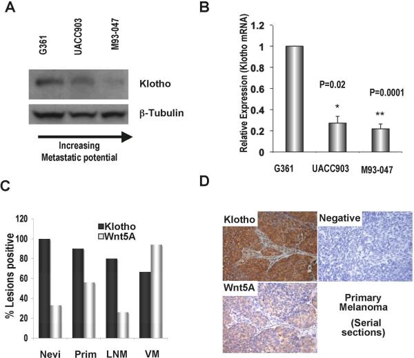
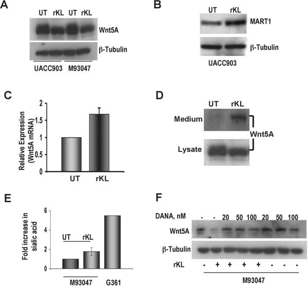
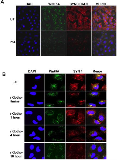
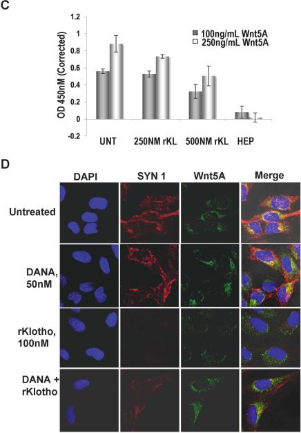
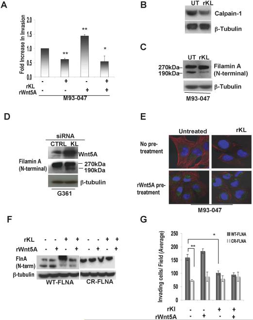
We have previously shown that Wnt5A-mediated signaling can promote melanoma metastasis. It has been shown that Wnt signaling is antagonized by the protein Klotho, which has been implicated in aging. We show here that in melanoma cells, expressions of Wnt5A and Klotho are inversely correlated. In the presence of recombinant Klotho (rKlotho), we show that Wnt5A internalization and signaling is decreased in high Wnt5A-expressing cells. Moreover, in the presence of rKlotho, we observe an increase in Wnt5A remaining in the medium, coincident with an increase in sialidase activity, and decrease in syndecan expression. These effects can be inhibited using a sialidase inhibitor. In addition to its effects on Wnt5A internalization, we also demonstrate that Klotho decreases melanoma cell invasive potential by a second mechanism that involves the inhibition of calpain and a resultant decrease in filamin cleavage, which we demonstrate is critical for melanoma cell motility.


Figures

Figure 1
Figure 1. Melanoma cells express the protein Klotho and its expression decreases with increased metastatic potential
(A) Total protein was isolated from melanoma cell lines of different metastatic potential and Klotho protein expression was measured by western blot analysis. β-Tubulin was used as a loading control. (B) Klotho mRNA expression by real-time PCR in G361, UACC903, and M93-047 melanoma cell lines normalized to 18S levels (n=4, error bars are standard deviation, *= p< 0.05, ** = p<0.005). (C) Percentages of lesions expressing Klotho, n= no of tumors per group. Prim= Primary melanoma, VM= Visceral metastases, LNM= Lymph node metastases. (D) Serial sections of primary melanoma stained for both Klotho and Wnt5A expression.
Figure 2
Figure 2. Wnt5A decreases Klotho expression
The expression and localization of Klotho (red) was examined in melanoma cell lines expressing different levels of Wnt5A using immunofluorescent confocal microscopy. (A) The expression of Klotho was low in M93-047 and UACC903 cell lines and (B) was abrogated in G361 cells following treatment with recombinant Wnt5A for 16 hours. (C) Total protein lysates from G361 cells were analyzed by Western blot for the expression of Klotho, which was decreased following treatment with recombinant Wnt5A for 16 hrs. β-Tubulin was used as a loading control. (D) Analysis of Klotho mRNA expression measured by real-time PCR analysis shows that treatment of G361 cells with recombinant Wnt5A decreased Klotho expression (n=3, error bars are standard error of the mean, * = p<0.05). Transfection of Wnt5A-expressing M93-047 with Wnt5A siRNA for 48 hrs resulted in an increase in (E) Klotho mRNA expression (Klotho mRNA expression was normalized to 18S levels (n=3, error bars are standard error of the mean, ** = p<0.005)), as well as (F) Klotho protein expression.
Figure 3
Figure 3. Klotho decreases Wnt5A expression
Total protein lysates from UACC903 and/or M93-047 cell lines were used to analyze Wnt5A and MART-1 protein expressions following treatment with recombinant Klotho for 16 hrs. (A) Wnt5A expression was decreased in both cell lines following rKlotho treatment. (B) MART-1 protein expression was increased in UACC903 following rKlotho treatment. β-Tubulin was used as a loading control. (C) Real-time PCR analysis of Wnt5A mRNA in M93-047 cells. Wnt5A expression was not significantly decreased in the presence of rKlotho. mRNA expression for Klotho was normalized to 18S levels (n=3, error bars are standard error of the mean). (D) Cells were treated with rKlotho for 16h, and the medium of the cells was examined for Wnt5A release. In the presence of Klotho, Wnt5A accumulates in the medium (upper panel), which correlates with a decrease in Wnt5A expression in total protein lysate (bottom panel). (E) Following appropriate treatment of cells, culture medium was collected and analyzed for sialidase activity as measured by sialic acid release. (Each sample was assayed in triplicate, n=3, error bars are standard error of the mean, * = p<0.05). (F) M93047 cells were treated for 16 hrs with recombinant Klotho in the presence or absence of DANA and total protein lysates were used to analyze Wnt5A protein expression. In the presence of DANA, the Klotho-induced decrease in Wnt5A expression is no longer observed.
Figure 4
Figure 4. Klotho prevents HSPG-mediated Wnt5A uptake and signaling
The expression and localization of Syndecan-1 (red) and Wnt5A (green) were examined in M93-047 cells using immunofluorescent confocal microscopy. (A) Treatment of M93-047 cells with recombinant Klotho for 16hrs shows that the decrease in Wnt5A expression in the presence of Klotho correlates with a decrease in the expression of Syndecan-1. (B) Co-localization of Wnt5A and syndecan-1 is affected as early as 5 mins after adding recombinant Klotho. (C) A solid phase binding assay demonstrates that in the presence of rKlotho, the binding of rWnt5A to rSyndecan is inhibited in a dose-dependent manner. (D) In the presence of DANA (50nM), treatment with recombinant klotho for 16 hrs no longer affects Wnt5A and syndecan expression levels as well as their co-localization.
Figure 5
Figure 5. Klotho negatively regulates the Wnt5A-mediated Filamin A cleavage and invasive potential
(A) M93-047 cells were subjected to a matrigel invasion assay. Invasion was measured by counting the cells in the bottom well 48 hrs after seeding on the matrigel-coated transwell filter. For each well, cells were counted in four different fields and averaged. The results are the average of three separate assays in which each condition was performed in triplicate. Error bars are standard error of the mean, * = p<0.05, **=p<0.005. (B) Calpain-1 protein expression was measured by Western blot analysis and was decreased in the presence of Klotho. (C) Total protein lysates from M93-047 cells were analyzed by Western blot for the presence of Filamin A cleavage. The N-terminal Filamin A cleavage fragment was identified as a 190 kDa band, and cleavage was decreased in the presence of Klotho. β-Tubulin was used as a loading control. (D) Expression of the N-terminal Filamin A cleavage fragment was increased in G361 cells transfected with Klotho SiRNA (E) The effect of excess rWnt5A on the Klotho-mediated expression and localization of filamin was examined in M93-047 cells. (F) M2 cells were transfected with either wildtype (WT) or Calpain resistant (CR) Filamin A (FLNA) treated with recombinant Klotho and /or recombinant Wnt5A for 16 hrs. (G) Total protein lysates were analyzed for the expression of N-terminal Filamin A cleavage and, (H) the invasive potential of the cells was measured using a matrigel invasion assay as described in (A). Error bars are standard error of the mean, * = p<0.05, **=p<0.005.

References

    1. BAUDRY M, DUBRIN R, BEASLEY L, LEON M, LYNCH G. Low levels of calpain activity in Chiroptera brain: implications for mechanisms of aging. Neurobiol Aging. 1986;7:255–8. - PubMed
    1. BESSE S, DELCAYRE C, CHEVALIER B, HARDOUIN S, HEYMES C, BOURGEOIS F, MOALIC JM, SWYNGHEDAUW B. Is the senescent heart overloaded and already failing? Cardiovasc Drugs Ther. 1994;8:581–7. - PubMed
    1. BLALOCK EM, PORTER NM, LANDFIELD PW. Decreased G-protein-mediated regulation and shift in calcium channel types with age in hippocampal cultures. J Neurosci. 1999;19:8674–84. - PMC - PubMed
    1. CAMPISI J, D'ADDA DI FAGAGNA F. Cellular senescence: when bad things happen to good cells. Nat Rev Mol Cell Biol. 2007;8:729–40. - PubMed
    1. CHA SK, ORTEGA B, KUROSU H, ROSENBLATT KP, KURO OM, HUANG CL. Removal of sialic acid involving Klotho causes cell-surface retention of TRPV5 channel via binding to galectin-1. Proc Natl Acad Sci U S A. 2008;105:9805–10. - PMC - PubMed

Publication types

MeSH terms