

The neurovascular unit, matrix proteases, and innate inflammation
- PMID: 20955425
- PMCID: PMC4547552
- DOI: 10.1111/j.1749-6632.2010.05760.x
The neurovascular unit, matrix proteases, and innate inflammation
Abstract
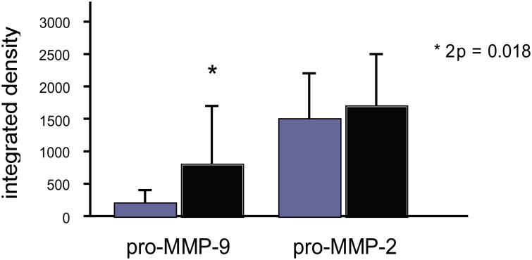
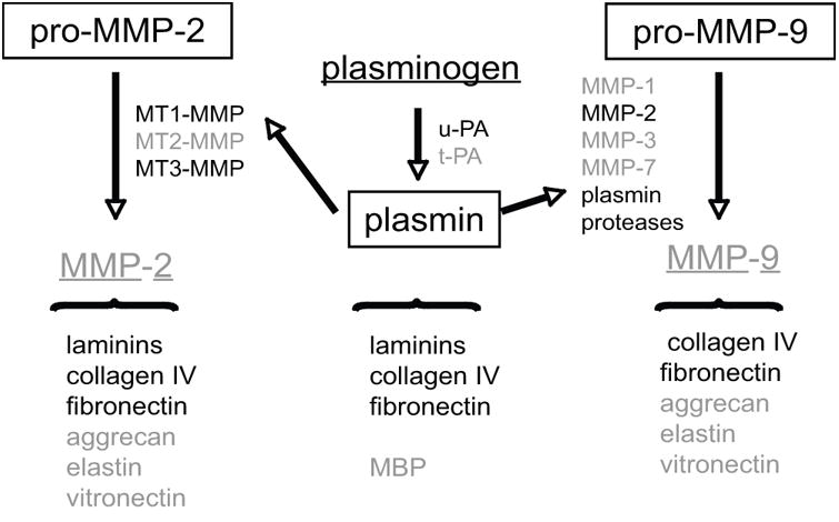
In the central nervous system, microvessel-neuron interactions appear highly coordinated. The rapid simultaneous responses of the microvasculature, neurons, and glia to focal ischemia in experimental ischemic stroke suggest that these responses could be viewed in a unitary fashion, rather than as individual components. The "neurovascular unit" consists of microvessels (endothelial cells-basal lamina matrix-astrocyte end-feet [and pericytes]), astrocytes, neurons and their axons, and other supporting cells that are likely to modulate the function of the "unit." Each cell component generates an inflammatory response to ischemia. Matrix metalloproteinase (MMP)-9 was first associated with hemorrhagic transformation following focal ischemia in an experimental model. A series of studies of ischemic stroke patients also suggests a relationship between MMP-9 levels and several consequences of ischemic injury, including hemorrhagic transformation. Recent experimental work suggests specific cell sources for MMP-9 generation and for matrix proteases from four distinct families that could impact neurovascular unit integrity.
© 2010 New York Academy of Sciences.
Conflict of interest statement
Figures
References
-
- del Zoppo GJ, et al. Polymorphonuclear leukocytes occlude capillaries following middle cerebral artery occlusion and reperfusion in baboons. Stroke. 1991;22:1276–1284. - PubMed
-
- Heo JH, et al. Matrix metalloproteinases increase very early during experimental focal cerebral ischemia. J Cereb Blood Flow Metab. 1999;19:624–633. - PubMed
-
- Okada Y, et al. P-selectin and intercellular adhesion molecule-1 expression after focal brain ischemia and reperfusion. Stroke. 1994;25:202–211. - PubMed
-
- Pitzer JE, del Zoppo GJ, Schmid-Schönbein GW. Neutrophil activation in smokers. Biorheology. 1996;33:45–58. - PubMed
-
- Hosomi N, et al. Rapid differential endogenous plasminogen activator expression after acute middle cerebral artery occlusion. Stroke. 2001;32:1341–1348. - PubMed
Publication types
MeSH terms
Substances
Grants and funding
LinkOut - more resources
Full Text Sources
Miscellaneous
