. 2010 Nov 2;107(44):18880-5.
doi: 10.1073/pnas.1005574107. Epub 2010 Oct 18.

Autophagy protein microtubule-associated protein 1 light chain-3B (LC3B) activates extrinsic apoptosis during cigarette smoke-induced emphysema

Affiliations

Autophagy protein microtubule-associated protein 1 light chain-3B (LC3B) activates extrinsic apoptosis during cigarette smoke-induced emphysema

Zhi-Hua Chen et al. Proc Natl Acad Sci U S A. .

Abstract

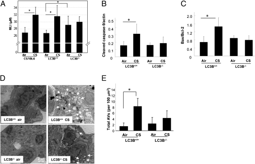
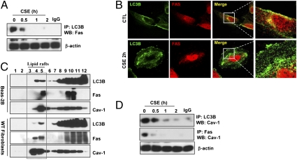
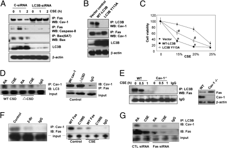
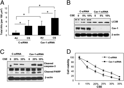
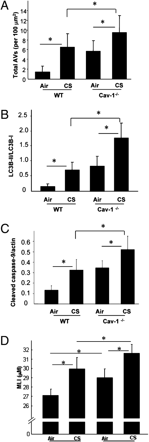
Chronic obstructive pulmonary disease (COPD) is a debilitating disease caused by chronic exposure to cigarette smoke (CS), which involves airway obstruction and alveolar loss (i.e., emphysema). The mechanisms of COPD pathogenesis remain unclear. Our previous studies demonstrated elevated autophagy in human COPD lung, and as a cellular and tissue response to CS exposure in an experimental model of emphysema in vivo. We identified the autophagic protein microtubule-associated protein 1 light chain-3B (LC3B) as a positive regulator of CS-induced lung epithelial cell death. We now extend these initial observations to explore the mechanism by which LC3B mediates CS-induced apoptosis and emphysema development in vivo. Here, we observed that LC3B(-/-) mice had significantly decreased levels of apoptosis in the lungs after CS exposure, and displayed resistance to CS-induced airspace enlargement, relative to WT littermate mice. We found that LC3B associated with the extrinsic apoptotic factor Fas in lipid rafts in an interaction mediated by caveolin-1 (Cav-1). The siRNA-dependent knockdown of Cav-1 sensitized epithelial cells to CS-induced apoptosis, as evidenced by enhanced death-inducing signaling complex formation and caspase activation. Furthermore, Cav-1(-/-) mice exhibited higher levels of autophagy and apoptosis in the lung in response to chronic CS exposure in vivo. In conclusion, we demonstrate a pivotal role for the autophagic protein LC3B in CS-induced apoptosis and emphysema, suggestive of novel therapeutic targets for COPD treatment. This study also introduces a mechanism by which LC3B, through interactions with Cav-1 and Fas, can regulate apoptosis.


Conflict of interest statement

The authors declare no conflict of interest.

Figures

Fig. 1.
Fig. 1.
LC3B regulates CS-induced autophagy, apoptosis, and emphysematous airspace enlargement in vivo. WT C57BL/6, LC3B+/+, or LC3B−/− mice were exposed to chronic CS exposure or air for 3 mo (C57BL/6, n = 9 for air, n = 8 for CS; LC3B+/+, n = 6 for air, n = 9 for CS; LC3B−/−, n = 9 for air, n = 9 for CS). (A) Quantification of MLIs (*P < 0.01). Representative images of mouse lungs with H&E staining are shown in Fig. S1. Western blot analysis and corresponding quantification for cleaved caspase-9 (B) and Bax/Bcl-2 ratio (C) in CS-exposed mouse lungs. Data are means ± SD; N.S., not significant; *P < 0.05. (D) Representative EM of mouse lung sections. Arrows indicate autophagic vacuoles (AVs). (E) EM images scored for number of AVs. The data are represented as AVs per 100 μm2; n = 20 images representative of each group; *P < 0.05.
Fig. 2.
Fig. 2.
LC3B-Cav-1-Fas interactions regulate CSE-induced autophagic and apoptotic cell death. (A) CSE-disrupted the LC3B-Fas interaction. Beas-2B cells were treated with CSE (10%) for the indicated times and their lysates were immunoprecipitated (IP) with anti-LC3B and Western immunoblotted (WB) for Fas. Cellular β-actin served as the standard. (B) Representative immunofluorescence images of LC3B and Fas staining in Beas-2B cells. Colocalization is indicated by the merged images (Right). (C) LC3B and Fas partially localize in lipid rafts. Beas-2B cells or WT fibroblasts were fractionated by sucrose-gradient ultracentrifugation and the fractions were immunoblotted for Fas, LC3B, and Cav-1. (D) CSE-disrupted the LC3B-Cav-1 and Fas-Cav-1 interaction. Cells were exposed to CSE (10%) for the indicated times, and their lysates were subjected to IP with anti-LC3B or Fas, and analyzed by WB for Cav-1. β-Actin served as the standard.
Fig. 3.
Fig. 3.
LC3B regulates Fas-mediated apoptosis in CSE-treated Beas-2B cells through binding interactions with Cav-1 and Fas. (A) Beas-2B cells were pretreated with control siRNA (C-siRNA) or LC3B-siRNA for 48 h, followed by CSE (10%) treatment for the indicated times. The lysates were then subjected to IP and WB analysis as indicated. (B) Mutation of LC3B at Y113 modulates the LC3B-Cav-1-Fas interactions under basal conditions. Beas-2B cells were transfected with WT or Y113A LC3B for 48 h, and the cell lysates were then subjected for IP and WB analysis as indicated. (C) Beas-2B cells were transfected with WT LC3B or mutant Y113A LC3B for 48 h, followed by the CSE treatment for an additional 24 h. Cells were then subjected to the 3-(4,5-dimethylthiazol-2-yl)-2,5-diphenyl tetrazolium bromide cell viability assay. Data represent mean ± SD; *P < 0.05 vs. corresponding vector control values; #P < 0.05 vs. corresponding values for WT-LC3B overexpression. (D) WT cav-1 and ΔCSD expression clones were transfected into Beas2B cells; 36 h after transfection, cells were exposed to CSE (10%). After 1 h, cell lysates were immunoprecipitated with anti-Cav-1 rabbit polyclonal antibodies. After separation on SDS/PAGE, respective mouse monoclonal antibodies were used to detect the interactions between Cav-1 and LC3B or Cav-1 and Fas, as indicated. The figure is representative of at least three independent experiments. (E) Cell lysates from 10% CSE-treated WT or Cav-1−/− fibroblasts were subjected to IP by LC3B and WB for Fas. A nonspecific IgG band served as the standard. The expression of Cav-1 and Fas in Cav-1−/− fibroblasts were assessed by Western analysis (Right), using β-actin as the standard. (F) Fas-Cav-1 interaction depends on palmitoylation domains. (Left) Beas2B cells were pretreated for 1 h with 100 μM 2-bromopalmitate (2-Br) or DMSO as a control. Cell lysates were subjected to coimmunoprecipitation assays. The figure is representative of at least three independent experiments. (Right) WT Fas and C199S mutant expression clones were transfected into Beas2B cells; 36 h after transfection, cells were exposed to CSE (10%). After 1 h, cell lysates were immunoprecipated with anti-Cav-1 or anti-Fas rabbit polyclonal antibodies. After fractionation on SDS/PAGE, respective mouse monoclonal antibodies were used as primary antibodies to detect interaction between Cav-1 and Fas. The figure is representative of at least three independent experiments. (G) Beas-2B cells were pretreated with control siRNA (C-siRNA) or Fas-siRNA for 48 h, followed by CSE (10%) treatment for the indicated times. The lysates were subjected to immunoprecipitation (IP) and Western blot (WB) analysis as indicated.
Fig. 4.
Fig. 4.
Cav-1 suppresses CSE-induced autophagy and apoptotic cell death in Beas-2B cells. (AC) Cells were transfected with siRNA for 48 h, followed by CSE treatment (10%) for an additional 24 h. (A) EM images of cells from each treatment group were scored for the number of AVs. The data are represented as AVs per 100 μm2; n = 20 images representative of each group. Data represent mean ± SD; *P < 0.05. (B) WB analysis of LC3B and Cav-1. β-actin served as standard. (C) Increased apoptosis in Cav-1-siRNA infected Beas-2B cells by CSE. Cells were transfected with siRNA for 48 h, and followed by CSE treatment at the indicated concentrations for an additional 24 h. Cell lysates were then analyzed for cleaved caspase-3 and cleaved poly(ADP ribose) polymerase. β-Actin served as the standard. (D) Cav-1 protects epithelial cells from CSE-induced cell death. Beas-2B cells treated with Cav-1-siRNA were exposed to CSE as indicated for 24 h, and cell viability was measured.
Fig. 5.
Fig. 5.
Cav-1 regulates CS-induced autophagy and apoptosis in vivo. WT C57BL/6 or Cav-1−/− mice were exposed to chronic CS exposure for 3 mo (each group, n = 5). (A) EM images were scored for number of AVs. The data are represented as AVs per 100 μm2; n = 20 images representative of each group. Western blot analysis and corresponding quantification for the LC3B-II/LC3B-I ratio (B) and cleaved caspase-9 (C) in CS-exposed mouse lungs. (D) MLI scoring of mouse lungs exposed to CS. Data represent mean ± SD; *P < 0.05. Representative images of mouse lungs with H&E staining are shown in Fig. S5.

References

    1. Lopez AD, Murray CC. The global burden of disease, 1990-2020. Nat Med. 1998;4:1241–1243. - PubMed
    1. Rabe KF, et al. Global Initiative for Chronic Obstructive Lung Disease. Global strategy for the diagnosis, management, and prevention of chronic obstructive pulmonary disease: GOLD executive summary. Am J Respir Crit Care Med. 2007;176:532–555. - PubMed
    1. Yoshida T, Tuder RM. Pathobiology of cigarette smoke-induced chronic obstructive pulmonary disease. Physiol Rev. 2007;87:1047–1082. - PubMed
    1. Petrache I, et al. Ceramide upregulation causes pulmonary cell apoptosis and emphysema-like disease in mice. Nat Med. 2005;11:491–498. - PMC - PubMed
    1. Kasahara Y, et al. Inhibition of VEGF receptors causes lung cell apoptosis and emphysema. J Clin Invest. 2000;106:1311–1319. - PMC - PubMed

Publication types

MeSH terms