. 2010 Nov 2;107(44):19126-31.
doi: 10.1073/pnas.1013032107. Epub 2010 Oct 18.

Neuronal Abelson helper integration site-1 (Ahi1) deficiency in mice alters TrkB signaling with a depressive phenotype

Affiliations

Neuronal Abelson helper integration site-1 (Ahi1) deficiency in mice alters TrkB signaling with a depressive phenotype

Xingshun Xu et al. Proc Natl Acad Sci U S A. .

Abstract

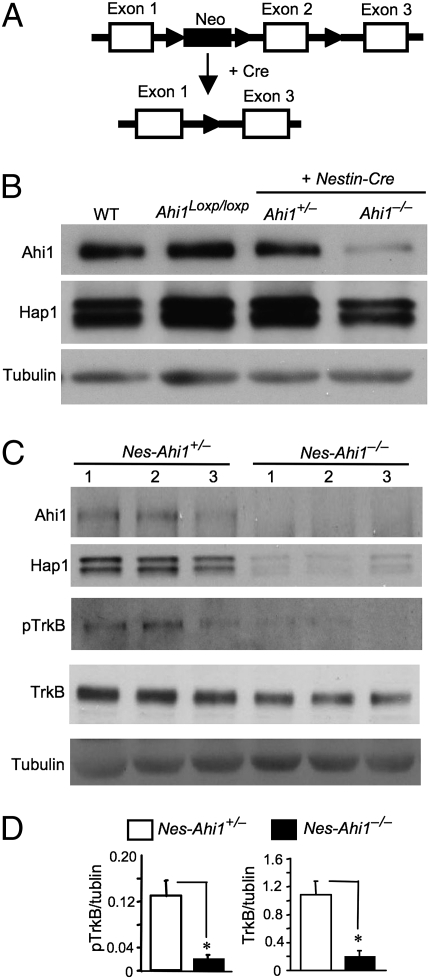
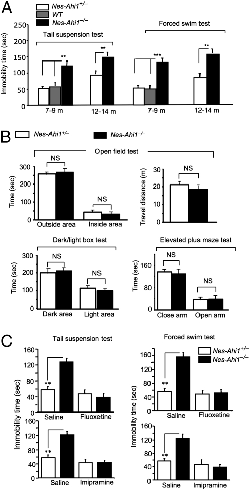
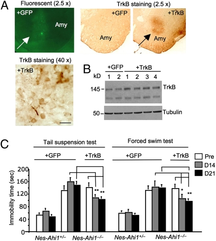
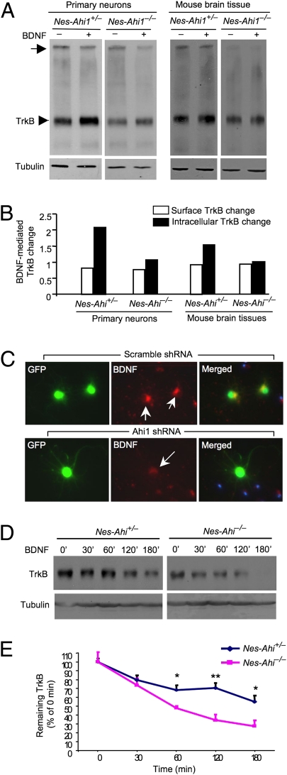
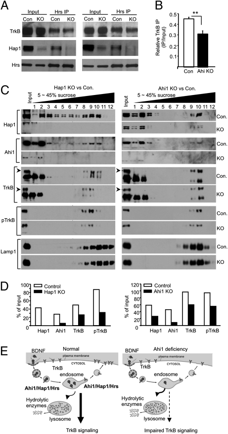
Recent studies suggest that the human Abelson helper integration site-1 (AHI1) gene on chromosome 6 is associated with susceptibility to schizophrenia and autism, two common neuropsychological disorders with depression symptoms. Mouse Ahi1 protein is abundant in the hypothalamus and amygdala, which are important brain regions for controlling emotion. However, the neuronal function of Ahi1 remains unclear. With the Cre-loxP system, we created a mouse model that selectively reduces Ahi1 expression in neuronal cells. Mice with neuronal Ahi1 deficiency show reduced TrkB level in the brain and depressive phenotypes, which can be alleviated by antidepressant drugs or by overexpression of TrkB in the amygdala. Ahi1 deficiency promotes the degradation of endocytic TrkB and reduces TrkB signaling in neuronal cells. Our findings suggest that impaired endocytic sorting and increased degradation of TrkB can induce depression and that this impaired pathway may serve as a previously uncharacterized therapeutic target for depression.


Conflict of interest statement

The authors declare no conflict of interest.

Figures

Fig. 1.
Fig. 1.
Ahi1 deficiency diminishes Hap1 and TrkB. (A) Generation of conditional Ahi1 knockout mice. Ahi1 exon 2 was removed by the Cre–loxP system to suppress Ahi1 expression. (B) Western blot analysis of Ahi1 and Hap1 expression in the hypothalamic tissue from wild-type (WT), floxed Ahi1 (Ahi1loxp/loxp), heterozygous (nes-Ahi1+/−), and homozygous conditional knockout (nes-Ahi1−/−) mice. (C) Total TrkB and pTrkB in the hypothalamus of control (nes-Ahi1+/−) and Ahi1-deficient (nes-Ahi1−/−) mice were examined by Western blotting. The blots were also probed with antibodies to Ahi1, Hap1, and tubulin. (D) Densitometric analysis of the ratios of TrkB or pTrkB to tubulin showing a decrease of total TrkB and pTrkB in Ahi1-deficient (nes-Ahi1−/−) mice (n = 3 each group, *P < 0.05).
Fig. 2.
Fig. 2.
Ahi1 deficiency leads to depressive behaviors. (A) Depressive phenotypes of nes-Ahi1−/− mice at 7–9 and 12–15 mo of age. *P < 0.05, **P < 0.01 between age-matched nes-Ahi1−/− and nes-Ahi1+/− or wild-type (WT) groups. Each group contains 8–16 mice. (B) Anxiety tests of conditional Ahi1 knockout mice. The open field test was performed, and time spent in the outside area and inside area of the cage (Upper Left) as well as distance traveled (Upper Right) were calculated. The times spent in the dark area and light area (Lower Left) and in the closed arm and open arm in the elevated plus maze test (Lower Right) were also recorded. NS, not significant or P > 0.05, n = 10. (C) The increased immobility time in TSTs and FSTs in the Ahi1-deficient mice (nes-Ahi1−/−) was reversed by the antidepressants imipramine (30 mg/kg) or fluoxetine (20 mg/kg), which was i.p. injected into mice for 30 min. The control is the injection of saline. *P < 0.05 or **P < 0.01.
Fig. 3.
Fig. 3.
Overexpression of TrkB in the amygdala of Ahi1-deficient mice partially rescues their depressive phenotype. (A) Stereotaxic injection of lentiviral GFP or TrkB into the amygdala (Amy) of mice resulted in the expression of GFP or TrkB in the injected area, which was revealed by fluorescent or TrkB immunostained (2.5× and 40×) micrographs. The antibody to TrkB (80G2, Cell Signaling Technology) did not react with endogenous mouse TrkB but was able to recognize overexpressed rat TrkB via lentiviral vector expression. (B) Western blotting showing the increased level of TrkB in four different amygdala tissues that had been injected with lentiviral TrkB for 2 weeks compared with two lentiviral GFP-injected amygdala tissues. (C) Stereotaxic injection of lentiviral TrkB reduced the immobility of Ahi1-deficient (nes-Ahi1−/−) mice in TSTs and FSTs, compared with those injected with lentiviral GFP. *P < 0.05 or **P < 0.01. Each group contains 8–12 mice. Mice were examined before (pre) and at 14 (D14) or 21 (D21) d after the lentiviral injection.
Fig. 4.
Fig. 4.
Ahi1 deficiency reduces the internalized TrkB and facilitates TrkB degradation. (A) Western blotting and BS3 crosslinking of membrane-bound receptors showing a reduction in membrane TrkB (arrow) and intracellular TrkB (arrowhead) in cultured primary brainstem cells and brainstem sections from nes-Ahi1−/− mice. BDNF (100 ng/mL for 1 h) was used to trigger TrkB internalization. (B) Quantifying BDNF-mediated changes of TrkB relative to that before BDNF treatment. (C) Decreased internalization of BDNF in cultured mouse brainstem cells treated with adenoviral Ahi1 shRNA. Arrows indicate neurons that coexpress adenoviral Ahi1 shRNA and GFP. (D) Cultured brainstem cells from control (nes-Ahi1+/−) mice and Ahi1-deficient (nes-Ahi1−/−) mice were treated with BDNF to induce TrkB internalization. TrkB Western blotting was then performed to assess the levels of TrkB at different times. (E) Quantitative analysis of the degradation of TrkB in brainstem slices of nes-Ahi1+/− and nes-Ahi1−/− mice. The ratios of TrkB to tubulin were measured via densitometry. The relative TrkB expression level at different times is expressed as percentage of the TrkB/tubulin ratio at 0 min before BDNF-induced internalization. The data were obtained from three independent experiments. *P < 0.05.
Fig. 5.
Fig. 5.
Ahi1 deficiency decreases the association of Hrs with TrkB and levels of TrkB in lysosomal fractions. (A) Hrs immunoprecipitation of brainstem tissues treated with BDNF (100 ng/mL for 30 min) and isolated from control (nes-Ahi1+/− or Con.) mice and Ahi1-deficient (nes-Ahi1−/− or KO) mice. Two immunoprecipitation results are presented with the one (Right) that used more input from nes-Ahi1−/− (KO) sample. Note that in the KO samples, there is reduced Hap1 in the input and precipitate samples, as well as decreased TrkB in the precipitate. (B) Quantification of the relative immunoprecipitated TrkB (ratios of precipitated to input TrkB) from three immunoprecipitation experiments. *P < 0.05. (C) Distribution of Hap1, Ahi1, TrkB, pTrkB, and lysosomal marker protein Lamp1 in 5–45% sucrose gradient fractions. Brain cortex tissues from Hap1 null (KO) and wild-type (Con.) mouse at P1–P2 (Left) or from (nes-Ahi1+/− or Con.) and Ahi1-deficient (nes-Ahi1−/− or KO) mice at 3 mo of age were analyzed. (D) Quantification of the amounts of proteins relative to input (% of input) in the lysosomal fractions (fraction-9 for Hap1 KO vs. control and fraction-10 for Ahi1 KO vs. control) isolated via sucrose gradient density centrifugation. (E) Proposed molecular mechanism for the depressive phenotype in Ahi1-deficient mice. BDNF triggers the internalization of TrkB receptor and signaling mediated by endocytic BDNF/TrkB. Ahi1/Hap1/Hrs complex is involved in intracellular trafficking of endocytic TrkB, prevents its degradation by lysosomes, and promotes its recycling to the plasma surface. When Ahi1 is reduced, more endocytic TrkB is transferred into lysosomes for degradation, resulting in the decreased level of TrkB and impaired TrkB signaling.

References

    1. Poirier Y, Jolicoeur P. Distinct helper virus requirements for Abelson murine leukemia virus-induced pre-B- and T-cell lymphomas. J Virol. 1989;63:2088–2098. - PMC - PubMed
    1. Ferland RJ, et al. Abnormal cerebellar development and axonal decussation due to mutations in AHI1 in Joubert syndrome. Nat Genet. 2004;36:1008–1013. - PubMed
    1. Dixon-Salazar T, et al. Mutations in the AHI1 gene, encoding jouberin, cause Joubert syndrome with cortical polymicrogyria. Am J Hum Genet. 2004;75:979–987. - PMC - PubMed
    1. Levi A, et al. Fine mapping of a schizophrenia susceptibility locus at chromosome 6q23: Increased evidence for linkage and reduced linkage interval. Eur J Hum Genet. 2005;13:763–771. - PubMed
    1. Torri F, et al. Fine mapping of AHI1 as a schizophrenia susceptibility gene: From association to evolutionary evidence. FASEB J. 2010;24:3066–3082. - PubMed

Publication types

MeSH terms