

Sildenafil reverses cardiac dysfunction in the mdx mouse model of Duchenne muscular dystrophy
- PMID: 20956307
- PMCID: PMC2973894
- DOI: 10.1073/pnas.1013077107
Sildenafil reverses cardiac dysfunction in the mdx mouse model of Duchenne muscular dystrophy
Abstract
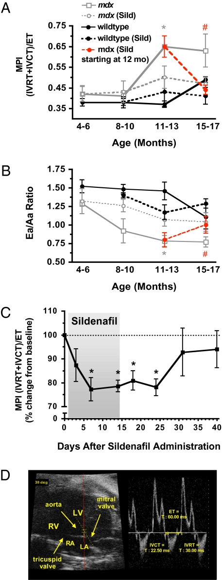
Duchenne muscular dystrophy (DMD) is a progressive and fatal genetic disorder of muscle degeneration. Patients with DMD lack expression of the protein dystrophin as a result of mutations in the X-linked dystrophin gene. The loss of dystrophin leads to severe skeletal muscle pathologies as well as cardiomyopathy, which manifests as congestive heart failure and arrhythmias. Like humans, dystrophin-deficient mice (mdx mice) show cardiac dysfunction as evidenced by a decrease in diastolic function followed by systolic dysfunction later in life. We have investigated whether sildenafil citrate (Viagra), a phosphodiesterase 5 (PDE5) inhibitor, can be used to ameliorate the age-related cardiac dysfunction present in the mdx mice. By using echocardiography, we show that chronic sildenafil treatment reduces functional deficits in the cardiac performance of aged mdx mice, with no effect on normal cardiac function in WT controls. More importantly, when sildenafil treatment was started after cardiomyopathy had developed, the established symptoms were rapidly reversed within a few days. It is recognized that PDE5 inhibitors can have cardioprotective effects in other models of cardiac damage, but the present study reports a prevention and reversal of pathological cardiac dysfunction as measured by functional analysis in a mouse model of DMD. Overall, the data suggest that PDE5 inhibitors may be a useful treatment for the cardiomyopathy affecting patients with DMD at early and late stages of the disease.
Conflict of interest statement
The authors declare no conflict of interest.
Figures


References
-
- Finsterer J, Stöllberger C. The heart in human dystrophinopathies. Cardiology. 2003;99:1–19. - PubMed
-
- Adamo CM. Evaluation of the Therapeutic Utility of Phosphodiesterase 5A Inhibition in the mdx Mouse Model of Duchenne Muscular Dystrophy. In: Schmidt HHW, Hofmann F, Stasch JP, editors. Handbook of Experimental Pharmacology: Phosphodiesterases as Drug Targets. Vol. 192. New York: Springer-Verlag; 2010. in press. - PMC - PubMed
-
- McNally EM. Duchenne muscular dystrophy: how bad is the heart? Heart. 2008;94:976–977. - PubMed
-
- Bushby K, Muntoni F, Bourke JP. 107th ENMC international workshop: The management of cardiac involvement in muscular dystrophy and myotonic dystrophy. 7th-9th June 2002, Naarden, the Netherlands. Neuromuscul Disord. 2003;13:166–172. - PubMed
Publication types
MeSH terms
Substances
Grants and funding
LinkOut - more resources
Full Text Sources
Other Literature Sources
Medical
Molecular Biology Databases
