

Endothelial histamine H1 receptor signaling reduces blood-brain barrier permeability and susceptibility to autoimmune encephalomyelitis
- PMID: 20956310
- PMCID: PMC2973887
- DOI: 10.1073/pnas.1008816107
Endothelial histamine H1 receptor signaling reduces blood-brain barrier permeability and susceptibility to autoimmune encephalomyelitis
Abstract
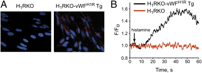
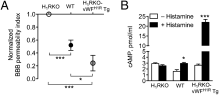
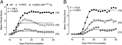
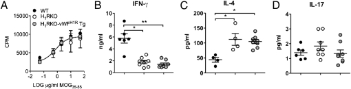
Disruption of the blood-brain barrier (BBB) underlies the development of experimental autoimmune encephalomyelitis (EAE) and multiple sclerosis. Environmental factors, such as Bordetella pertussis, are thought to sensitize central endothelium to biogenic amines like histamine, thereby leading to increased BBB permeability. B. pertussis-induced histamine sensitization (Bphs) is a monogenic intermediate phenotype of EAE controlled by histamine H(1) receptor (Hrh1/H(1)R). Here, we transgenically overexpressed H(1)R in endothelial cells of Hrh1-KO (H(1)RKO) mice to test the role of endothelial H(1)R directly in Bphs and EAE. Unexpectedly, transgenic H(1)RKO mice expressing endothelial H(1)R under control of the von Willebrand factor promoter (H(1)RKO-vWF(H1R) Tg) were Bphs-resistant. Moreover, H(1)RKO-vWF(H1R) Tg mice exhibited decreased BBB permeability and enhanced protection from EAE compared with H(1)RKO mice. Thus, contrary to prevailing assumptions, our results show that endothelial H(1)R expression reduces BBB permeability, suggesting that endothelial H(1)R signaling may be important in the maintenance of cerebrovascular integrity.
Conflict of interest statement
The authors declare no conflict of interest.
Figures
Similar articles
-
von-Willebrand factor influences blood brain barrier permeability and brain inflammation in experimental allergic encephalomyelitis.Am J Pathol. 2008 Sep;173(3):892-900. doi: 10.2353/ajpath.2008.080001. Epub 2008 Aug 7. Am J Pathol. 2008. PMID: 18688020 Free PMC article.
-
H(1)R expression by CD11B(+) cells is not required for susceptibility to experimental allergic encephalomyelitis.Cell Immunol. 2012 Jul-Aug;278(1-2):27-34. doi: 10.1016/j.cellimm.2012.06.012. Epub 2012 Jul 14. Cell Immunol. 2012. PMID: 23121973 Free PMC article.
-
Systemic lack of canonical histamine receptor signaling results in increased resistance to autoimmune encephalomyelitis.J Immunol. 2013 Jul 15;191(2):614-22. doi: 10.4049/jimmunol.1203137. Epub 2013 Jun 14. J Immunol. 2013. PMID: 23772030 Free PMC article.
-
Identification of Bphs, an autoimmune disease locus, as histamine receptor H1.Science. 2002 Jul 26;297(5581):620-3. doi: 10.1126/science.1072810. Science. 2002. PMID: 12142541
-
The action of TH17 cells on blood brain barrier in multiple sclerosis and experimental autoimmune encephalomyelitis.Hum Immunol. 2020 May;81(5):237-243. doi: 10.1016/j.humimm.2020.02.009. Epub 2020 Feb 28. Hum Immunol. 2020. PMID: 32122685 Review.
Cited by
-
Elevated CSF histamine levels in multiple sclerosis patients.Fluids Barriers CNS. 2013 May 9;10:19. doi: 10.1186/2045-8118-10-19. eCollection 2013. Fluids Barriers CNS. 2013. PMID: 23659456 Free PMC article.
-
A genetic locus complements resistance to Bordetella pertussis-induced histamine sensitization.Commun Biol. 2023 Mar 6;6(1):244. doi: 10.1038/s42003-023-04603-w. Commun Biol. 2023. PMID: 36879097 Free PMC article.
-
The allergy mediator histamine confers resistance to immunotherapy in cancer patients via activation of the macrophage histamine receptor H1.Cancer Cell. 2022 Jan 10;40(1):36-52.e9. doi: 10.1016/j.ccell.2021.11.002. Epub 2021 Nov 24. Cancer Cell. 2022. PMID: 34822775 Free PMC article.
-
Combinatorial roles for histamine H1-H2 and H3-H4 receptors in autoimmune inflammatory disease of the central nervous system.Eur J Immunol. 2012 Jun;42(6):1536-46. doi: 10.1002/eji.201141859. Eur J Immunol. 2012. PMID: 22678907 Free PMC article.
-
Endothelial Adora2a Activation Promotes Blood-Brain Barrier Breakdown and Cognitive Impairment in Mice with Diet-Induced Insulin Resistance.J Neurosci. 2019 May 22;39(21):4179-4192. doi: 10.1523/JNEUROSCI.2506-18.2019. Epub 2019 Mar 18. J Neurosci. 2019. PMID: 30886019 Free PMC article.
References
Publication types
MeSH terms
Substances
Grants and funding
- NS069628/NS/NINDS NIH HHS/United States
- HL089243/HL/NHLBI NIH HHS/United States
- NS060901/NS/NINDS NIH HHS/United States
- R01 NS036526/NS/NINDS NIH HHS/United States
- R01 AI058052/AI/NIAID NIH HHS/United States
- AI041747/AI/NIAID NIH HHS/United States
- R01 NS060901/NS/NINDS NIH HHS/United States
- AI058052/AI/NIAID NIH HHS/United States
- P01 AI045666/AI/NIAID NIH HHS/United States
- HL44455/HL/NHLBI NIH HHS/United States
- NS036526/NS/NINDS NIH HHS/United States
- NS061014/NS/NINDS NIH HHS/United States
- R01 NS069628/NS/NINDS NIH HHS/United States
- AI045666/AI/NIAID NIH HHS/United States
- R01 HL044455/HL/NHLBI NIH HHS/United States
- R01 AI041747/AI/NIAID NIH HHS/United States
- R01 NS061014/NS/NINDS NIH HHS/United States
LinkOut - more resources
Full Text Sources
Molecular Biology Databases
Research Materials
Miscellaneous
