. 2010 Nov 2;107(44):18950-5.
doi: 10.1073/pnas.1011036107. Epub 2010 Oct 18.

Neutralization of West Nile virus by cross-linking of its surface proteins with Fab fragments of the human monoclonal antibody CR4354

Affiliations

Neutralization of West Nile virus by cross-linking of its surface proteins with Fab fragments of the human monoclonal antibody CR4354

Bärbel Kaufmann et al. Proc Natl Acad Sci U S A. .

Abstract

Many flaviviruses are significant human pathogens, with the humoral immune response playing an essential role in restricting infection and disease. CR4354, a human monoclonal antibody isolated from a patient, neutralizes West Nile virus (WNV) infection at a postattachment stage in the viral life-cycle. Here, we determined the structure of WNV complexed with Fab fragments of CR4354 using cryoelectron microscopy. The outer glycoprotein shell of a mature WNV particle is formed by 30 rafts of three homodimers of the viral surface protein E. CR4354 binds to a discontinuous epitope formed by protein segments from two neighboring E molecules, but does not cause any detectable structural disturbance on the viral surface. The epitope occurs at two independent positions within an icosahedral asymmetric unit, resulting in 120 binding sites on the viral surface. The cross-linking of the six E monomers within one raft by four CR4354 Fab fragments suggests that the antibody neutralizes WNV by blocking the pH-induced rearrangement of the E protein required for virus fusion with the endosomal membrane.


Conflict of interest statement

The authors declare no conflict of interest.

Figures

Fig. 1.
Fig. 1.
CryoEM reconstruction of WNV in complex with Fab fragments of the neutralizing human MAb CR4354. (A) Surface rendering of the 3D image reconstruction of WNV (green) in complex with Fab CR4354 (blue) at 13.7-Å resolution, viewed down an icosahedral twofold axis. The black triangle outlines an ASU. (B) Central cross-section of the 3D image reconstruction of the complex viewed down an icosahedral twofold axis. The positions of icosahedral two-, three-, and fivefold axes are indicated. (Scale bar, 100 Å.)
Fig. 2.
Fig. 2.
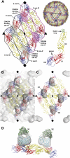
Binding pattern of CR4354 relative to the E glycoprotein on the surface of mature WNV. (A) Arrangement of the fitted E glycoproteins on the viral surface. DI, DII, and DIII of each E monomer are colored red, yellow, and blue, respectively. The fusion loop is shown in green and N-linked glycans on DI in magenta. The gray outlines highlight the three different E monomers (E mol-A, -B, and -C) of one ASU. The typical raft structure of three almost parallel E homodimers (Left) is formed by the three E monomers of one ASU with the E molecules of a twofold symmetry-related ASU (E mol-A′, -B′, and -C′; magenta outlines). Gray shadows indicate the footprints of the two independent CR4354 Fab binding sites, X1 and X2, per ASU. Pink shadows show the symmetry-related Fab binding sites (X1 Vogt and X2 Vogt) in the neighboring ASU. (B) Fit of three independent E molecules per ASU (E mol-A through -C) into the cryoEM density of the WNV–CR4354 complex. The twofold symmetry-related E molecules (E mol-A′ through -C′) are also shown. The positions of the icosahedral five-, three-, and twofold symmetry axes are marked with symbols (pentagons, triangles, and ovals, respectively). At the chosen contour level for the complex density (2.8σ), only the variable domain density of the Fab is visible. (C) Difference density (gray) between the WNV–Fab complex and WNV superpositioned onto the E protein raft, viewed down an icosahedral twofold axis. The difference map is contoured as the complex density in B. CR4354 Fab binds to two independent sites per ASU, X1 and X2. Fab binding to X1 cross-links two E monomers of the ASU, whereas binding to X2 locks the arrangement of E in rafts by cross-linking symmetry-related E molecules. (D) Side view of the fitted Fab CR4354 at positions X1 and X2. The variable domains of the Fabs are shown in cyan, the constant domains in green. The difference map is contoured at 1.4σ of the complex density. Molecular graphics images were produced using Chimera (45).
Fig. 3.
Fig. 3.
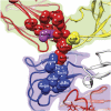
Detailed view of the CR4354 epitope at binding site X1 (for binding site X2, see Fig. S3). The two independent E molecules that contribute to the CR4354 epitope are shown as ribbon diagrams and highlighted by background shading (E mol-A, green; -B, purple). The individual E domains are color-coded as follows: DI, red; DII, yellow; DIII, blue. The atoms of the E residues that constitute the CR4354 epitope are depicted as balls. Residue Lys136, previously identified as part of the epitope by neutralization escape mutant analysis (21), is shown in magenta.
Fig. 4.
Fig. 4.
The cross-linking of E molecules within a raft by CR4354 Fab fragments. Shown are the six E molecules of two twofold-related ASUs (A, B, C and A′, B′, C′) forming a raft. The position of the icosahedral twofold symmetry axis is indicated. E monomers that are engaged by a Fab are opaque. The Fab footprints at sites X1 and X2 and the twofold-related sites X1′ and X2′ are superpositioned onto the E raft. (A) CR4354 Fabs bound to site X1 and its twofold symmetry-related position X1′, only, engage two E molecules of one ASU each (A + B and A′ + B′, respectively). As indicated by molecule separation and arrows, this scenario possibly allows the dissociation of the central dimer upon low pH exposure. (B) CR4354 Fabs bound to site X2 and X2′, only, cross-link two E monomers from two neighboring ASUs each (C + A′ and C′ + A, respectively). As indicated by molecule separation and arrows, this scenario possibly allows the dissociation of the outer dimers upon low pH exposure. (C) Utilization of X1 and X2 results in cross-linking of all six E monomers within a raft and prohibits the dissociation of the E glycoprotein rafts before forming a fusogenic state.

Similar articles

Cited by

References

    1. Petersen LR, Hayes EB. West Nile virus in the Americas. Med Clin North Am. 2008;92:1307–1322, ix. - PubMed
    1. Rey FA, Heinz FX, Mandl C, Kunz C, Harrison SC. The envelope glycoprotein from tick-borne encephalitis virus at 2 A resolution. Nature. 1995;375:291–298. - PubMed
    1. Modis Y, Ogata S, Clements D, Harrison SC. A ligand-binding pocket in the dengue virus envelope glycoprotein. Proc Natl Acad Sci USA. 2003;100:6986–6991. - PMC - PubMed
    1. Nybakken GE, Nelson CA, Chen BR, Diamond MS, Fremont DH. Crystal structure of the West Nile virus envelope glycoprotein. J Virol. 2006;80:11467–11474. - PMC - PubMed
    1. Kanai R, et al. Crystal structure of West Nile virus envelope glycoprotein reveals viral surface epitopes. J Virol. 2006;80:11000–11008. - PMC - PubMed

Publication types

MeSH terms

LinkOut - more resources