

T-cell receptor-driven lymphomagenesis in mice derived from a reprogrammed T cell
- PMID: 20956329
- PMCID: PMC2973852
- DOI: 10.1073/pnas.1013230107
T-cell receptor-driven lymphomagenesis in mice derived from a reprogrammed T cell
Abstract
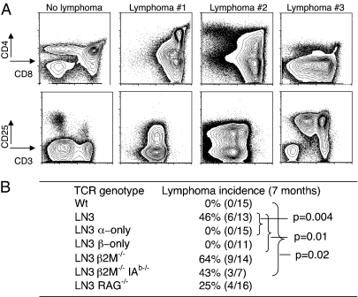
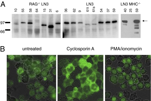
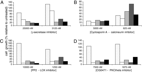
The conversion of mature somatic cells into pluripotent stem cells, both by nuclear transfer and transduction with specific "reprogramming" genes, represents a major advance in regenerative medicine. Pluripotent stem cell lines can now be generated from an individual's own cells, facilitating the generation of immunologically acceptable stem cell-based therapeutics. Many cell types can undergo nuclear reprogramming, leading to the question of whether the identity of the reprogrammed cell of origin has a biological consequence. Peripheral blood, containing a mixture of T, B, NK, and myeloid cell types, represents one potential source of reprogrammable cells. In this study, we describe the unique case of mice derived from a reprogrammed T cell. These mice have prerearranged T-cell receptor (TCR) genes in all cells. Surprisingly, ≈50% of mice with prerearranged TCR genes develop spontaneous T cell lymphomas, which originate in the thymus. The lymphomas arise from developing T cells, and contain activated Notch1, similar to most human and mouse T-cell acute lymphoblastic lymphomas. Furthermore, lymphomagenesis requires the expression of both prerearranged TCRα and TCRβ genes, indicating a critical role for TCR signaling. Furthermore, inhibitors of multiple branches of TCR signaling suppress lymphoma growth, implicating TCR signaling as an essential component in lymphoma proliferation. The lymphomagenesis in mice derived from a reprogrammed T cell demonstrates the deleterious consequences of misregulation of the TCR rearrangement and signaling pathways and illustrates one case of cellular reprogramming where the identity of the cell of origin has profound consequences.
Conflict of interest statement
Conflict of interest statement: J.H. and J.S. are founding members of Complegen, Inc.
Figures
Similar articles
-
Oncogenesis of T-ALL and nonmalignant consequences of overexpressing intracellular NOTCH1.J Exp Med. 2008 Nov 24;205(12):2851-61. doi: 10.1084/jem.20081561. Epub 2008 Nov 3. J Exp Med. 2008. PMID: 18981238 Free PMC article.
-
Anaplastic large cell lymphoma arises in thymocytes and requires transient TCR expression for thymic egress.Nat Commun. 2016 Jan 12;7:10087. doi: 10.1038/ncomms10087. Nat Commun. 2016. PMID: 26753883 Free PMC article.
-
Bi-Allelic TCRα or β Recombination Enhances T Cell Development but Is Dispensable for Antigen Responses and Experimental Autoimmune Encephalomyelitis.PLoS One. 2015 Dec 22;10(12):e0145762. doi: 10.1371/journal.pone.0145762. eCollection 2015. PLoS One. 2015. PMID: 26693713 Free PMC article.
-
Synergy between the pre-T cell receptor and Notch: cementing the alphabeta lineage choice.J Exp Med. 2006 Oct 2;203(10):2233-7. doi: 10.1084/jem.20060998. Epub 2006 Sep 25. J Exp Med. 2006. PMID: 17000868 Free PMC article. Review.
-
[Analyses of the rearrangement of T-cell receptor- and immunoglobulin genes in the diagnosis of lymphoproliferative disorders].Veroff Pathol. 1995;144:1-109. Veroff Pathol. 1995. PMID: 7856305 Review. German.
Cited by
-
Adoptive Transfer of CD8+ T Cells Generated from Induced Pluripotent Stem Cells Triggers Regressions of Large Tumors Along with Immunological Memory.Cancer Res. 2016 Jun 15;76(12):3473-83. doi: 10.1158/0008-5472.CAN-15-1742. Epub 2016 Apr 12. Cancer Res. 2016. PMID: 27197199 Free PMC article.
-
Reappraisal of nodal Epstein-Barr Virus-negative cytotoxic T-cell lymphoma: Identification of indolent CD5+ diseases.Cancer Sci. 2018 Aug;109(8):2599-2610. doi: 10.1111/cas.13652. Epub 2018 Jun 26. Cancer Sci. 2018. PMID: 29845715 Free PMC article.
-
Endogenous dendritic cells from the tumor microenvironment support T-ALL growth via IGF1R activation.Proc Natl Acad Sci U S A. 2016 Feb 23;113(8):E1016-25. doi: 10.1073/pnas.1520245113. Epub 2016 Feb 9. Proc Natl Acad Sci U S A. 2016. PMID: 26862168 Free PMC article.
-
Generation of integration-free human induced pluripotent stem cells from postnatal blood mononuclear cells by plasmid vector expression.Nat Protoc. 2012 Nov;7(11):2013-21. doi: 10.1038/nprot.2012.121. Epub 2012 Oct 18. Nat Protoc. 2012. PMID: 23080273 Free PMC article.
-
Pluripotent stem cell-based cancer therapy: promise and challenges.Sci Transl Med. 2012 Mar 28;4(127):127ps9. doi: 10.1126/scitranslmed.3003920. Sci Transl Med. 2012. PMID: 22461639 Free PMC article. Review.
References
-
- Takahashi K, Yamanaka S. Induction of pluripotent stem cells from mouse embryonic and adult fibroblast cultures by defined factors. Cell. 2006;126:663–676. - PubMed
-
- Hochedlinger K, Jaenisch R. Monoclonal mice generated by nuclear transfer from mature B and T donor cells. Nature. 2002;415:1035–1038. - PubMed
-
- Kim JB, et al. Direct reprogramming of human neural stem cells by OCT4. Nature. 2009;461:649–653. - PubMed
-
- Aoi T, et al. Generation of pluripotent stem cells from adult mouse liver and stomach cells. Science. 2008;321:699–702. - PubMed
Publication types
MeSH terms
Substances
Grants and funding
LinkOut - more resources
Full Text Sources
Other Literature Sources
Molecular Biology Databases
