. 2010 Nov 2;107(44):18838-43.
doi: 10.1073/pnas.1007387107. Epub 2010 Oct 18.

Keap1 perceives stress via three sensors for the endogenous signaling molecules nitric oxide, zinc, and alkenals

Affiliations

Keap1 perceives stress via three sensors for the endogenous signaling molecules nitric oxide, zinc, and alkenals

Michael McMahon et al. Proc Natl Acad Sci U S A. .

Abstract

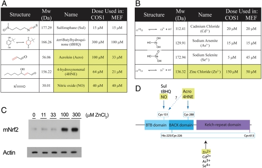
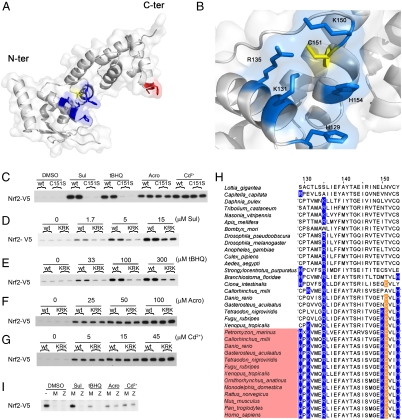
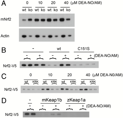
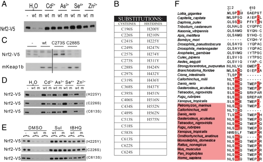
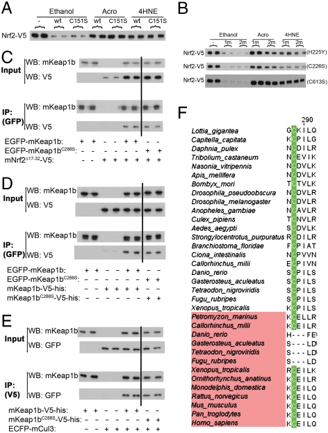
Recognition and repair of cellular damage is crucial if organisms are to survive harmful environmental conditions. In mammals, the Keap1 protein orchestrates this response, but how it perceives adverse circumstances is not fully understood. Herein, we implicate NO, Zn(2+), and alkenals, endogenously occurring chemicals whose concentrations increase during stress, in this process. By combining molecular modeling with phylogenetic, chemical, and functional analyses, we show that Keap1 directly recognizes NO, Zn(2+), and alkenals through three distinct sensors. The C288 alkenal sensor is of ancient origin, having evolved in a common ancestor of bilaterans. The Zn(2+) sensor minimally comprises H225, C226, and C613. The most recent sensor, the NO sensor, emerged coincident with an expansion of the NOS gene family in vertebrates. It comprises a cluster of basic amino acids (H129, K131, R135, K150, and H154) that facilitate S-nitrosation of C151. Taken together, our data suggest that Keap1 is a specialized sensor that quantifies stress by monitoring the intracellular concentrations of NO, Zn(2+), and alkenals, which collectively serve as second messengers that may signify danger and/or damage.


Conflict of interest statement

The authors declare no conflict of interest.

Figures

Fig. 1.
Fig. 1.
Chemical analysis of mKeap1b. Structures and concentrations of electrophiles (A) and inorganic compounds (B) used to treat cells. The chemical tBHQ is shown in equilibrium with its quinone derivative. Endogenously occurring chemicals are highlighted in yellow. (C) Samples prepared from mouse embryonic fibroblasts treated with the indicated doses of ZnCl2 for 2 h were blotted for Nrf2 and actin. D, A cartoon of mKeap1b indicating its BTB, BACK and Kelch domains, the three sensors identified in this paper, and the chemicals that trigger them. Although it is likely that Acro and 4HNE trigger the C151-based sensor, this conjecture remains to be formally proven.
Fig. 2.
Fig. 2.
Characterization of the C151-based sensor. (A) Homology model of amino acids 59–240 of mKeap1b. (B) A scaled-up view of C151 (yellow) highlighting its juxtaposition with H129, K131, R135, K150, and H154 (blue). (CG) A plasmid expressing wild-type mKeap1b (WT), mKeap1bC151S (C151S), or mKeap1b bearing triple K131, R135, and K150 to Met substitutions (KRK) was cotransfected into COS1 cells with plasmids expressing mNrf2-V5 and β-galactosidase (β-gal) (a control for transfection efficiency). Blotting for mNrf2-V5 was performed 2 h after chemical treatment. The blots shown are typical of n≥3 independent experiments. (H) Sequence alignment of Keap1 homologues highlighting the amino acid signature of the C151-based sensor. Keap1b paralogues are named in pink. The remaining sequences are of Keap1a proteins. (I) An empty plasmid (−) or one expressing either mKeap1b (M) or zKeap1a (Z) was cotransfected into Keap1b-/- mouse embryonic fibroblast cells with plasmids expressing mNrf2-V5 and β-gal. Blotting for mNrf2-V5 was performed 2 h after chemical treatment (n = 2 independent experiments).
Fig. 3.
Fig. 3.
NO is an endogenous trigger of the C151-based sensor. (A) WT or Keap1b-/- (ko) mouse embryonic fibroblast (MEF) cells were exposed to DEA-NO/AM for 2 h before blotting for Nrf2 or actin. (B and C) A plasmid expressing mKeap1b (WT), mKeap1C151S (C151S), or mKeap1b bearing triple K131, R135 and K150 to Met substitutions (KRK) was cotransfected into COS1 cells with plasmids expressing mNrf2-V5 and β-galactosidase (β-gal). Cells were treated with DEA-NO/AM for 2 h before blotting for mNrf2-V5. (D) An empty plasmid (−) or one expressing mKeap1b or zKeap1a was cotransfected into Keap1b-/- MEF cells with plasmids expressing mNrf2-V5 and β-gal. Blotting for mNrf2-V5 was performed 2 h after DEA-NO/AM treatment. All blots are typical of n≥3 independent experiments, except for blot D where n = 2.
Fig. 4.
Fig. 4.
The Zn2+ sensor. (A) An empty plasmid (−) or one expressing mKeap1b (WT), or mKeap1bC151S (m) was cotransfected into COS1 cells with plasmids expressing mNrf2-V5 and β-galactosidase (β-gal). Blotting for mNrf2-V5 was performed 2 h after chemical treatment. (B) Amino acid substitutions introduced into the BACK and Kelch domains of mKeap1b. (C) An empty plasmid (−) or one expressing mKeap1b (WT), mKeap1bC273S (C273S), or mKeap1bC288S (C288S) was cotransfected into Keap1b-/- mouse embryonic fibroblast cells with plasmids expressing mNrf2-V5 and β-gal. Blotting was performed 24 h later. Two lanes have been cropped from the middle of these blots, indicated by the vertical line, as they are not relevant to this paper. (D and E) An empty plasmid (−) or one expressing either mKeap1b (WT), or a mutated form of mKeap1b (m) was cotransfected into COS1 cells with plasmids expressing mNrf2-V5 and β-gal. Cells were treated with chemicals for 2 h before blotting for the stated proteins. Three separate substitutions were evaluated (H225Y, C226S, and C613S). (F) The amino acid signature of the Zn2+ sensor is highlighted in red. All immunoblots shown are representative of n≥3 independent experiments.
Fig. 5.
Fig. 5.
The alkenal sensor. (A) An empty plasmid (−) or one expressing mKeap1b (WT), or mKeap1bC151S (C151S) was cotransfected into COS1 cells with plasmids expressing mNrf2-V5 and β-galactosidase (β-gal). Blotting for mNrf2-V5 was performed 2 h after treatment of cells with chemicals. (B) An empty plasmid (−) or one expressing mKeap1bC151S (m) or mKeap1bC151S bearing an additional second substitution (2 m) was cotransfected into COS1 cells with plasmids expressing mNrf2-V5 and β-gal. Blotting for mNrf2-V5 was performed after cells had been treated with the stated chemicals for 2 h; the second substitution utilized in each experiment is identified to the right of each blot. (CE) The indicated combinations of proteins were expressed in COS1 cells and EGFP-tagged or V5-tagged material was immunoprecipitated. Immunoprecipitates (IP) and input samples were blotted with the indicated antibodies. (F) C288 is highlighted in this sequence alignment. All immunoblots shown are representative of n≥3 independent experiments.

References

    1. Kultz D. Molecular and evolutionary basis of the cellular stress response. Annu Rev Physiol. 2005;67:225–257. - PubMed
    1. Gasch AP. Comparative genomics of the environmental stress response in ascomycete fungi. Yeast. 2007;24:961–976. - PubMed
    1. Sykiotis GP, Bohmann D. Keapl/Nrf2 signaling regulates oxidative stress tolerance and lifespan in Drosophila. Dev Cell. 2008;14:76–85. - PMC - PubMed
    1. Singh A, et al. Dysfunctional KEAP1-NRF2 interaction in non-small-cell lung cancer. PLoS Med. 2006;3:1865–1876. - PMC - PubMed
    1. Sykiotis GP, Bohmann D. Stress-activated cap‘n’collar transcription factors in aging and human disease. Sci Signal. 2010;3:1–22. - PMC - PubMed

Publication types

MeSH terms