. 2010 Nov 9;107(45):19213-8.
doi: 10.1073/pnas.1009645107. Epub 2010 Oct 18.

Engineering of a synthetic electron conduit in living cells

Affiliations

Engineering of a synthetic electron conduit in living cells

Heather M Jensen et al. Proc Natl Acad Sci U S A. .

Abstract

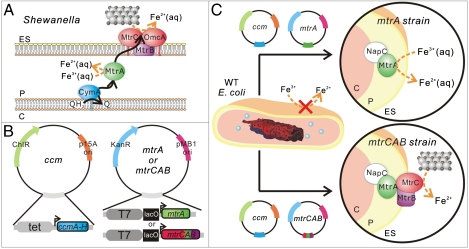
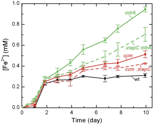
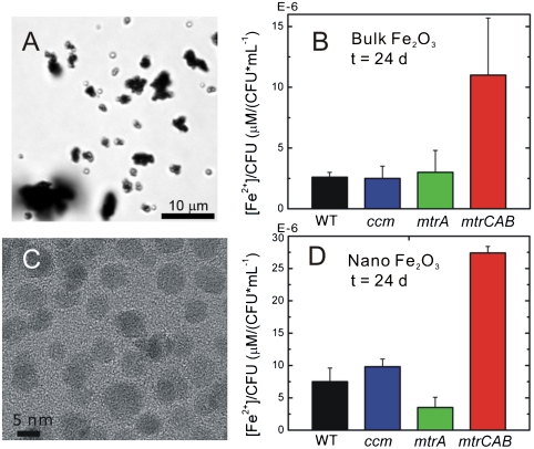
Engineering efficient, directional electronic communication between living and nonliving systems has the potential to combine the unique characteristics of both materials for advanced biotechnological applications. However, the cell membrane is designed by nature to be an insulator, restricting the flow of charged species; therefore, introducing a biocompatible pathway for transferring electrons across the membrane without disrupting the cell is a significant challenge. Here we describe a genetic strategy to move intracellular electrons to an inorganic extracellular acceptor along a molecularly defined route. To do so, we reconstitute a portion of the extracellular electron transfer chain of Shewanella oneidensis MR-1 into the model microbe Escherichia coli. This engineered E. coli can reduce metal ions and solid metal oxides ∼8× and ∼4× faster than its parental strain. We also find that metal oxide reduction is more efficient when the extracellular electron acceptor has nanoscale dimensions. This work demonstrates that a genetic cassette can create a conduit for electronic communication from living cells to inorganic materials, and it highlights the importance of matching the size scale of the protein donors to inorganic acceptors.


Conflict of interest statement

The authors declare no conflict of interest.

Figures

Fig. 1.
Fig. 1.
(A) Schematic of proposed extracellular electron transfer pathway in Shewanella oneidensis MR-1 where ES denotes the extracellular space, P denotes the periplasm, and C denotes the cytoplasm. The silver and black spheres represent extracellular iron oxide. (B) Schematic of plasmids used to create the ccm, mtrA, and mtrCAB strains in E. coli. (C) Schematic of the engineered mtrA and mtrCAB strains for soluble and extracellular metal reduction.
Fig. 2.
Fig. 2.
Expression of full-length redox-active MtrA and MtrC in E. coli. Heme-stained SDS-PAGE gels of (A) periplasmic fractions and (B) membrane fractions of the WT, ccm, mtrA, and mtrCAB strains. (C) Anti-MtrB immunoblot of membrane fractions of the WT, ccm, mtrA, and mtrCAB strains. (D) Absorption spectra of the periplasmic fraction of the mtrA strain under oxidizing and reducing conditions. (E) Absorption spectra of the membrane fraction of the mtrCAB strain under oxidizing and reducing conditions.
Fig. 3.
Fig. 3.
Reduction of 10 mM Fe(III) citrate to Fe(II) as a function of time for the WT, ccm, mtrA, and mtrCAB E. coli strains. Error bars represent the standard deviation between triplicates from separate starting cultures.
Fig. 4.
Fig. 4.
Direct link of MtrA redox state to Fe(III) citrate reduction. (A) Absorption spectra showing the α-band of MtrA in high-density, anaerobic cell suspensions of the mtrA strain before and after the addition of Fe(III) citrate. MtrA begins reduced (black line, strong α-band absorption), but is oxidized upon the addition of 50 μM Fe(III) citrate (red line). Over time, the α-band absorbance recovers (colored dotted lines). (B) ΔA552 nm and Fe(II) concentration immediately before and after Fe(III) citrate addition as measured by the ferrozine assay.
Fig. 5.
Fig. 5.
NapC is not the sole electron donor to MtrA. Reduction of 10 mM Fe(III) citrate to Fe(II) by WT, ccm, ΔnapC ccm, mtrA, and ΔnapC mtrA strains.
Fig. 6.
Fig. 6.
MtrCAB reduces solid α-Fe2O3. (A) Brightfield optical image of bulk α-Fe2O3, d ∼ 5 μm. (B) The concentration of bulk α-Fe2O3 reduced by WT, cmm, mtrA, mtrCAB strains normalized by colony forming units after 24 d. (C) Transmission electron microscopy of crystalline α-Fe2O3 nanoparticles, d = 13 nm. (B) The concentration of α-Fe2O3 nanoparticles reduced by WT, ccm, mtrA, mtrCAB strains normalized by colony forming units after 24 d.

References

    1. Patolsky F, et al. Detection, stimulation, and inhibition of neuronal signals with high-density nanowire transistor arrays. Science. 2006;313:1100–1104. - PubMed
    1. Ionescu-Zanetti C, et al. Mammalian electrophysiology on a microfluidic platform. Proc Natl Acad Sci USA. 2005;102:9112–9117. - PMC - PubMed
    1. Marsili E, et al. Shewanella secretes flavins that mediate extracellular electron transfer. Proc Natl Acad Sci USA. 2008;105:3968–3973. - PMC - PubMed
    1. Park DH, Zeikus JG. Electricity generation in microbial fuel cells using neutral red as an electronophore. Appl Environ Microbiol. 2000;66:1292–1297. - PMC - PubMed
    1. Gunawardena A, Fernando S, To F. Performance of a yeast-mediated biological fuel cell. Int J Mol Sci. 2008;9:1893–1907. - PMC - PubMed

Publication types

LinkOut - more resources