Comment
. 2010 Oct 18;191(2):233-6.
doi: 10.1083/jcb.201009048.

Cells in tight spaces: the role of cell shape in cell function

Affiliations
Comment

Cells in tight spaces: the role of cell shape in cell function

Jagesh V Shah. J Cell Biol. .

Abstract

In this issue, Pitaval et al. (2010. J. Cell Biol. doi:10.1083/jcb.201004003) demonstrate that cell geometry can regulate the elaboration of a primary cilium. Their findings and approaches are part of a historical line of inquiry investigating the role of cell shape in intracellular organization and cellular function.


Figures

Figure 1.
Figure 1.
Cell shape controls mitotic orientation. (A) Drawing by O. Hertwig of the confined frog embryo after its first division illustrating Hertwig’s rule. The image was scanned by Google Docs from Hertwig (1893). (B) Without apparent interactions with the cell boundary, we might expect the small mitotic spindle in a large cell to position randomly, but it orients according to Hertwig’s rule. This image was adapted from Wühr et al. (2009). (C and D) Micropatterned fibronectin in various geometries results in well-defined mitotic orientations (C) determined by actin-rich retraction fibers (D; green). blue, DNA; arrowheads, positions of spindle poles. (E) Computational modeling predicts the expected orientation based on adhesion pattern shape. C–E are reprinted from Théry et al., 2007 with permission from Nature Publishing Group.
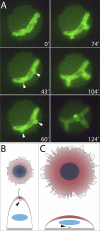
Figure 2.
Figure 2.
Cell shape controls cytoplasmic organization. (A) Time-lapse imaging of a deformed fission yeast cell in a microwell (30 µm in diameter) results in the local accumulation of bud6-3xEGFP, a polarity marker (43′, white arrowheads), and subsequent cell growth from the ectopic site (60′–124′; experiment performed in a cdc25-22 tea1Δ background). Reprinted from Minc et al., 2009 with permission from Elsevier. (B) Cellular confinement on a small extracellular matrix island results in low cortical tension (red), migration of the centriole pair (arrowhead) above the nucleus (blue), and the elaboration of a primary cilium. (C) Cell spreading on a larger island results in higher cortical tension (red), centriole trapping below the nucleus (arrowhead), and the inability to form a primary cilium. Adapted from Pitaval et al. (2010).

Comment on

Similar articles

Cited by

References

    1. Chen C.S., Mrksich M., Huang S., Whitesides G.M., Ingber D.E. 1997. Geometric control of cell life and death. Science. 276:1425–1428 10.1126/science.276.5317.1425 - DOI - PubMed
    1. Driesch H. 1893. Entwicklungsmechanische Studien. Zeitschrift für Wissenschaftliche Zoologie. 55:1–62
    1. Gerdes J.M., Davis E.E., Katsanis N. 2009. The vertebrate primary cilium in development, homeostasis, and disease. Cell. 137:32–45 10.1016/j.cell.2009.03.023 - DOI - PMC - PubMed
    1. Gibson W.T., Gibson M.C. 2009. Cell topology, geometry, and morphogenesis in proliferating epithelia. Curr. Top. Dev. Biol. 89:87–114 10.1016/S0070-2153(09)89004-2 - DOI - PubMed
    1. Grill S.W., Hyman A.A. 2005. Spindle positioning by cortical pulling forces. Dev. Cell. 8:461–465 10.1016/j.devcel.2005.03.014 - DOI - PubMed

Publication types