. 2010 Dec 17;285(51):40322-32.
doi: 10.1074/jbc.M110.137604. Epub 2010 Oct 18.

Sphingolipid-mediated inhibition of apoptotic cell clearance by alveolar macrophages

Affiliations

Sphingolipid-mediated inhibition of apoptotic cell clearance by alveolar macrophages

Daniela N Petrusca et al. J Biol Chem. .

Abstract

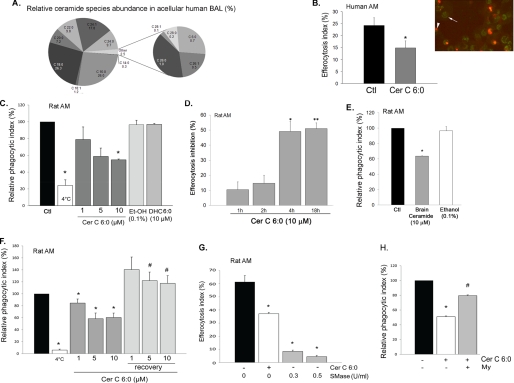
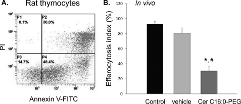
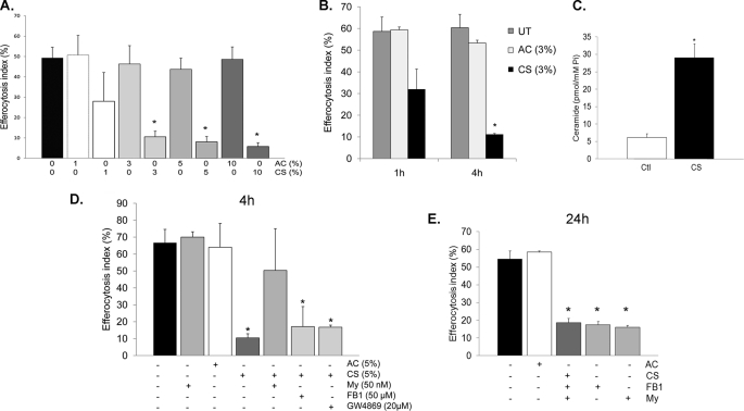
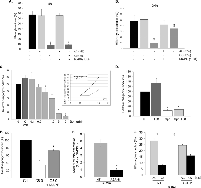
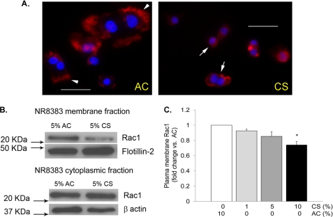
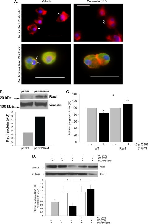
A decreased clearance of apoptotic cells (efferocytosis) by alveolar macrophages (AM) may contribute to inflammation in emphysema. The up-regulation of ceramides in response to cigarette smoking (CS) has been linked to AM accumulation and increased detection of apoptotic alveolar epithelial and endothelial cells in lung parenchyma. We hypothesized that ceramides inhibit the AM phagocytosis of apoptotic cells. Release of endogenous ceramides via sphingomyelinase or exogenous ceramide treatments dose-dependently impaired apoptotic Jurkat cell phagocytosis by primary rat or human AM, irrespective of the molecular species of ceramide. Similarly, in vivo augmentation of lung ceramides via intratracheal instillation in rats significantly decreased the engulfment of instilled target apoptotic thymocytes by resident AM. The mechanism of ceramide-induced efferocytosis impairment was dependent on generation of sphingosine via ceramidase. Sphingosine treatment recapitulated the effects of ceramide, dose-dependently inhibiting apoptotic cell clearance. The effect of ceramide on efferocytosis was associated with decreased membrane ruffle formation and attenuated Rac1 plasma membrane recruitment. Constitutively active Rac1 overexpression rescued AM efferocytosis against the effects of ceramide. CS exposure significantly increased AM ceramides and recapitulated the effect of ceramides on Rac1 membrane recruitment in a sphingosine-dependent manner. Importantly, CS profoundly inhibited AM efferocytosis via ceramide-dependent sphingosine production. These results suggest that excessive lung ceramides may amplify lung injury in emphysema by causing both apoptosis of structural cells and inhibition of their clearance by AM.


Figures

FIGURE 1.
FIGURE 1.
Ceramide inhibits AM efferocytosis ex vivo. A, abundance of ceramide species measured in the human BAL acellular fluid by combined LC-MS/MS. The data shown are the averages of n = 15 BAL obtained from apparently healthy volunteers. B–F, engulfment efficiency of human (B) or rat AM was assessed after AM were pretreated with ceramides for 4 h (unless otherwise stated) and co-cultured for 1 h with PI-stained apoptotic Jurkat cells. B, quantification of human AM efferocytosis by microscopy. Right panel, representative image of AM exhibiting green auto-fluorescence (arrowhead) with internalized PI-stained apoptotic Jurkat cells (red, arrow). Left panel, efferocytosis index calculated as the percentage of AM with engulfed PI-stained cells relative to the total AM number, using coded slides (means ± S.E.; *, p < 0.05; Student's t test) after treatment with vehicle (Ctl) or ceramide (Cer 6:0; 10 μm; 4 h). C and D, dose dependence (C; at 4 h) and kinetics (D) of inhibitory effect of Cer 6:0, its vehicle ethanol (Et-OH), or its precursor DHC (6:0) on rat AM efferocytosis. C, efferocytosis was measured by flow cytometry and expressed as relative phagocytic index (percentage of efferocytosis of treated AM relative to that of untreated AM). White bar, AM co-incubated with apoptotic Jurkats at 4 °C (mean ± S.E.; n = 14; (*, p < 0.05 versus control). D, efferocytosis was measured by flow cytometry and expressed as inhibitory activity (%) compared with untreated AM (mean ± S.E.; n = 3; *, p = 0.007; **, p = 0.005 versus control). E, rat AM efferocytosis was measured by flow cytometry and expressed as relative phagocytic index (percentage of untreated control: black bar) following treatment with brain ceramides (a porcine extract containing long chain ceramides) (10 μm; 4 h) or with vehicle (mean ± S.E.; n = 3; *, p < 0.05 versus control). F, effect of post-ceramide recovery of rat AM efferocytosis determined by flow cytometry and expressed as relative phagocytic index (percentage of untreated AM). Rat primary AM were treated with ceramide C6:0 (1, 5, or 10 μm; 4 h) and then allowed to recover overnight in regular growth medium prior to efferocytosis assay (means ± S.E.; n = 3; *, p < 0.05 versus control; #, p < 0.05 versus corresponding treatments). G, rat AM efferocytosis was measured by flow cytometry and expressed as efferocytosis index following treatment (4 h) with active neutral SMase at the indicated concentrations before challenge with apoptotic targets. Treatment with Cer 6:0 (10 μm; 4 h; white bar) is shown for comparison (means ± S.E.; n = 3; *, p < 0.05 versus control). H, rat AM efferocytosis expressed as relative phagocytic index (percentage of untreated control: black bar) following treatment with Cer 6:0 (10 μm; 4 h) after preincubation with myriocin, an inhibitor of SPT in the de novo ceramide synthesis pathway (My, 50 nm, 2 h; n = 3; means ± S.E.; *, p < 0.05 versus control; #, p < 0.05 versus Cer 6:0).
FIGURE 2.
FIGURE 2.
Ceramide inhibits AM efferocytosis in vivo. A, apoptosis of target thymocytes following ex vivo incubation (24 h), measured by flow cytometry following dual staining with PI and annexin V (representative flow panel; cells in the right upper and right lower panels are apoptotic). B, in vivo AM efferocytosis of intratracheally delivered PI-labeled apoptotic thymocytes (30 min), assessed by flow cytometry. AM were recovered by BAL from Sprague-Dawley rats treated intratracheally with either Cer 16:0 (PEG 2000-conjugated; 10 mg/kg; 24 h) or vehicle (PEG 2000); means ± S.E.; n = 4; *, p < 0.05 versus untreated; #, p < 0.05 versus vehicle).
FIGURE 3.
FIGURE 3.
CS inhibits AM efferocytosis in part by de novo ceramide synthesis via SPT. A, effect of aqueous extract of ambient AC or CS (1, 3, 5, and 10% v:v; 4 h) on rat AM efferocytosis, measured by flow cytometry (means ± S.E.; n = 10; *, p < 0.005 versus control). B, time-dependent effects of CS (3% CS extract v:v) on rat AM efferocytosis, measured by flow cytometry; means ± S.E.; n = 3; *, p < 0.05 versus untreated control (UT). C, total ceramides 24 h after removal of rat AM from treatment with CS (3%; 4 h), measured by tandem mass spectrometry, followed by normalization by intracellular inorganic phosphorus content; means ± S.E.; n = 4; *, p < 0.01 versus control. D and E, rat AM efferocytosis of PI-labeled apoptotic Jurkat cells following CS exposure (3–5%; 4 h) and specific ceramide synthesis inhibitors myriocin (My; 50 nm; 2 h), fumonisin (FB1; 5 μm; 2 h), or GW4869 (20 μm; 30 min). Engulfment was assessed after 4 h of CS exposure (D) or 24 h after removal of AM from the CS treatment (E) and quantified by flow cytometry (means ± S.E.; n = 3; *, p < 0.05 versus AC).
FIGURE 4.
FIGURE 4.
CS inhibition of AM efferocytosis is sphingosine-dependent. A and B, rat AM efferocytosis following inhibition of sphingosine synthesis with the ceramidase inhibitor (MAPP; 1 μm, 2 h) and exposure to CS (3%; 4 h) assessed immediately (A) or 24 h following removal of AM from the CS treatment (B) (means ± S.E.; *, p < 0.05 versus untreated control cells; #, p < 0.05 versus CS; n = 4). C, rat AM efferocytosis after treatment with sphingosine (Sph) at the indicated concentrations (4 h) or methanol vehicle (Veh; 0.7%; 4 h; means ± S.E.; n = 3; *, p < 0.001 versus untreated). Inset, inhibitory effect on efferocytosis (%) of sphingosine and S1P treatment at the indicated concentrations (4 h) on AM efferocytosis. D, rat AM efferocytosis following treatment (4 h) with ceramide synthase inhibitor fumonisin B1 (FB1; 5 μm; 2 h), sphingosine (3 μm), or sphingosine (3 μm) in the presence of FB1 (means ± S.E.; n = 3; *, p < 0.05 versus untreated (UT) control). E, AM efferocytosis after treatment with ceramide C8:0 (10 μm, 4 h) and the ceramidase inhibitor MAPP (1 μm, 2 h; mean ± S.E.; *, p < 0.05 versus untreated control; #, p < 0.05 versus C8:0; n = 3). F, acid ceramidase (ASAH1) mRNA expression (relative to GAPDH) measured in primary rat AM by real time PCR after 72 h of transient transfection with siRNA targeting ASAH1 (1 μm) or with nontarget siRNA (NT) (means ± S.D.; *, p < 0.005 versus NT siRNA; n = 2). G, rat AM efferocytosis following inhibition of sphingosine synthesis with acid ceramidase siRNA (1 μm, 72 h) and exposure to CS (3%; 4 h) or AC, assessed 24 h after removal of AM from the CS treatment (means ± S.E.; *, p < 0.05 versus control; #, p < 0.05 versus CS/NT; n = 2).
FIGURE 5.
FIGURE 5.
CS decreases Rac1 membrane abundance. A, representative fluorescence micrographs of AM (NR8383 cells) stained for actin (with Texas Red phalloidin; red) and nuclei (DAPI; blue) following treatment with AC or CS extract (5%; 4 h). Note that control cells (left panel) exhibit pronounced ruffling of the plasma membrane (arrows), whereas CS-treated cells (right panel) have markedly reduced membrane ruffle formation. Scale bar, 50 μm. B, Rac1 membrane abundance detected in protein lysates from total membrane and cytoplasmic fractions obtained from NR8383 cells treated with CS extract (5%; 4 h) or AC. The proteins were detected by Western blotting using a specific Rac1 antibody; flotillin-2 and β-actin were used as loading controls. C, densitometry of Rac1 expression detected by Western blotting normalized by loading control, expressed as fold change versus AC control (means ± S.E.; n = 3; *, p < 0.05 versus AC).
FIGURE 6.
FIGURE 6.
Ceramide inhibits AM efferocytosis through Rac1 down-regulation. A, representative fluorescence micrographs of AM (either alone in the upper panels or co-incubated with apoptotic Jurkat cells in the lower panels) stained for actin (with Texas Red phalloidin; red), nuclear marker (DAPI; blue), and Rac1 (with Rac1 antibody conjugated to Alexa fluor 488; green; lower panels only) following treatment with Cer C6:0 (10 μm; 4 h) or control vehicle (0.1% ethanol). Note that control cells exhibit ruffles of the plasma membrane (arrows), whereas ceramide-treated cells have a near loss of membrane ruffle formation (double arrowhead) and decreased Rac1 staining. The engulfed Jurkat apoptotic cells are seen in control cells (asterisk), surrounded by a Rac1-rich phagosome membrane (arrow). Scale bar, 50 μm. The values are representative of n = 2 experiments. B, Rac1 protein level in NR8383 cells transfected with constitutive active Rac1 or control plasmid. Vinculin immunoblot was used as loading control. C, phagocytic index of wild type NR8383 macrophages and those overexpressing Rac1 treated with ceramide (Cer C6:0; 10 μm; 4 h); note the lack of inhibitory effect of ceramide on efferocytosis in cells expressing a constitutively active Rac1 (means ± S.E.; n = 3). D, Rac1 plasma membrane abundance detected in protein lysates from total membrane fractions obtained from NR8383 cells treated with CS extract (3%; 4 h) or AC with or without a ceramidase inhibitor pretreatment (MAPP; 1 μm, 2 h). The proteins were detected by Western blotting using a specific Rac1 antibody, CD71 was used as loading control; densitometry of Rac1 expression was normalized by loading control (means ± S.E.; n = 3; *, p < 0.05).

Similar articles

Cited by

References

    1. Yoshida T., Tuder R. M. (2007) Physiol. Rev. 87, 1047–1082 - PubMed
    1. Kasahara Y., Tuder R. M., Taraseviciene-Stewart L., Le Cras T. D., Abman S., Hirth P. K., Waltenberger J., Voelkel N. F. (2000) J. Clin. Invest. 106, 1311–1319 - PMC - PubMed
    1. Tuder R. M., Petrache I., Elias J. A., Voelkel N. F., Henson P. M. (2003) Am. J. Respir. Cell Mol. Biol. 28, 551–554 - PubMed
    1. Tuder R. M., Zhen L., Cho C. Y., Taraseviciene-Stewart L., Kasahara Y., Salvemini D., Voelkel N. F., Flores S. C. (2003) Am. J. Respir. Cell Mol. Biol. 29, 88–97 - PubMed
    1. Rangasamy T., Cho C. Y., Thimmulappa R. K., Zhen L., Srisuma S. S., Kensler T. W., Yamamoto M., Petrache I., Tuder R. M., Biswal S. (2004) J. Clin. Invest. 114, 1248–1259 - PMC - PubMed

Publication types

MeSH terms