. 2010 Dec;30(24):5752-63.
doi: 10.1128/MCB.01147-10. Epub 2010 Oct 18.

Targeted inactivation of Mapk4 in mice reveals specific nonredundant functions of Erk3/Erk4 subfamily mitogen-activated protein kinases

Affiliations

Targeted inactivation of Mapk4 in mice reveals specific nonredundant functions of Erk3/Erk4 subfamily mitogen-activated protein kinases

Justine Rousseau et al. Mol Cell Biol. 2010 Dec.

Abstract

Erk4 and Erk3 are atypical members of the mitogen-activated protein (MAP) kinase family. The high sequence identity of Erk4 and Erk3 proteins and the similar organization of their genes imply that the two protein kinases are paralogs. Recently, we have shown that Erk3 function is essential for neonatal survival and critical for the establishment of fetal growth potential and pulmonary function. To investigate the specific functions of Erk4, we have generated mice with a targeted disruption of the Mapk4 gene. We show that Erk4-deficient mice are viable and fertile and exhibit no gross morphological or physiological anomalies. Loss of Erk4 is not compensated by changes in Erk3 expression or activity during embryogenesis or in adult tissues. We further demonstrate that additional loss of Erk4 does not exacerbate the fetal growth restriction and pulmonary immaturity phenotypes of Erk3(-/-) mice and does not compromise the viability of Erk3(+/-) neonates. Interestingly, behavioral phenotyping revealed that Erk4-deficient mice manifest depression-like behavior in the forced-swimming test. Our analysis indicates that the MAP kinase Erk4 is dispensable for mouse embryonic development and reveals that Erk3 and Erk4 have acquired specialized functions through evolutionary diversification.


Figures

FIG. 1.
FIG. 1.
Generation of Erk4-deficient mice. (A) Schematic representation of the Mapk4 locus, wild-type allele, targeting vector, and mutated allele. Exons 1 to 6 are represented by numbered gray or black bars or boxes. Gray bars or boxes represent untranslated regions (UTRs), and black bars or boxes represent coding regions. The targeting vector carries a neomycin resistance gene (Neo) and the GFP reporter gene fused in frame with the Mapk4 coding sequence initiation codon. A KpnI restriction site was inserted in the GFP gene to facilitate identification of the mutated allele. The positions of KpnI (K), BamHI (B), and EcoRI (E) restriction sites are shown. (B) Southern blot analysis of KpnI-digested genomic DNA from two correctly targeted ES clones (clones 117 and 148): 5′ probe, wild-type (Wt) allele (9.8 kb) and mutant allele (6.8 kb); 3′ probe, mutant allele (5.1 kb). Lanes: C, randomly integrated control; M, 1-kb marker. (C) PCR analysis of genomic DNA from 3-week-old Erk4+/− intercross progeny. (D) Immunoblot (IB) analysis of Erk4 protein expression in total cell extracts of Erk4+/+ and Erk4−/− mouse embryonic fibroblasts (MEFs).
FIG. 2.
FIG. 2.
Absence of Erk4 does not affect the proliferation rate of primary MEFs. (A) The proliferation rate of passage 3 (P3) MEFs prepared from Mapk4−/− or wild-type (WT) littermate embryos was measured by the MTT assay. Values are expressed as fold increase in cell number and correspond to the means ± standard errors of the means (SEMs) (error bars) of 7 or 8 independent MEF preparations. (B) Proliferation curves of MEFs isolated from Mapk4−/− embryos, Mapk4−/−; Mapk6−/− embryos, or wild-type embryos.
FIG. 3.
FIG. 3.
Absence of Erk4 is not compensated by changes in Erk3 expression or activity. (A and B) mRNA expression during embryonic development. Total RNA was extracted from wild-type or Erk4−/− whole embryos at different embryonic stages. Expression of Erk4 and Erk3 mRNA was analyzed by quantitative RT-PCR. (A) Levels of Erk3 and Erk4 mRNA in wild-type mice are expressed as fold difference relative to mRNA expression at E9.5 and represent the means plus SEMs (error bars) (n = 3). (B) Levels of Erk3 mRNA in Erk4−/− embryos are expressed relative to their expression levels in Erk4+/+ embryos at the corresponding embryonic day (E9.5 to E18.5). Values are means plus SEMs (n = 3). (C and D) Protein expression in adult tissues. Total proteins were extracted by homogenization of whole tissues from 8-week-old wild-type and Erk4−/− littermates, and equal amounts of proteins were resolved by SDS-polyacrylamide gel electrophoresis. Expression of Erk4 (C) and Erk3 (D) was analyzed by immunoblotting (IB) with specific antibodies. Cellular extracts of Erk3+/+ and Erk3−/− MEFs were used as control to show the specificity of Erk3 antibody. The specificity of anti-Erk4 antibody is presented in Fig. 1D. (E) Kinase activity. Protein extracts from Erk4+/+ and Erk4−/− brains were incubated for 4 h at 4°C with recombinant His6-MK5-GST. Erk3/Erk4-MK5 complexes were pulled down, and the phosphotransferase activity of MK5 was measured in a coupled assay using PRAK substrate peptide as the substrate. Results are expressed as fold change in activity and represent the means plus SEMs of 3 experiments. The values for kinase activity for Erk4+/+ and Erk4−/− mice are significantly different (P < 0.05) as indicated by the asterisk and bracket.
FIG. 4.
FIG. 4.
Distribution of Erk4 mRNA in adult mouse brain. (A) Expression of Erk4 mRNA was analyzed by in situ hybridization on mouse brain sagittal sections (from Lateral 2.04 mm to Lateral 0.96 mm, as referred to in the Paxinos and Franklin mouse brain atlas [25]). The specific expression pattern was assessed with a mix of five antisense probes targeting different regions of Erk4 mRNA. ACB, accumbens nucleus; CA1, CA1 field of the hippocampus; cbgl, cerebellar granular layer; CP, caudate putamen; Cx(LII), cortical layer 2; dcn, deep cerebellar nuclei; Epl, external plexiform layer of the olfactory bulb; grDG, granular layer of the dentate girus; GrO, granule cell layer of the olfactory bulb; icp, inferior cerebellar peduncle; int, internal capsule; LR4V, lateral recess of the fourth ventricle; LV, lateral ventricle; Pir, piriform cortex; Post, posterior subiculum; Pr5, principal sensory trigeminal nucleus; sp5, spinal trigeminal tract; Tu, olfactory tubercle. (B) Representative photographs of hybridization signal in sagittal sections from the brains of Erk4+/+ and Erk4−/− mice.
FIG. 5.
FIG. 5.
Loss of Erk4 does not impact on postnatal brain neurogenesis. (A) Quantification of BrdU- and double-cortin (DCX)-positive cells in Erk4+/+ and Erk4−/− dentate gyrus of 12-week-old mice. (B) Quantification of BrdU-positive granular and glomerular cells in Erk4+/+ and Erk4−/− olfactory bulb. Results are shown as means ± SEMs (n = 5 to 8). (C) Representative images of brain sections from Erk4+/+ and Erk4−/− mice stained with BrdU and DCX.
FIG. 6.
FIG. 6.
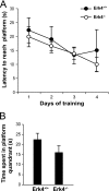
Normal spatial learning in Erk4-deficient mice. Spatial learning and memory performance were assessed by the Morris water maze test. (A) Time to reach the hidden platform during 4 days of training. (B) Time spent in the target platform quadrant during the probe session. Results are expressed as means ± SEMs (n = 5 to 7).
FIG. 7.
FIG. 7.
Erk4-deficient mice manifest depression-related behavior. Mice were analyzed for depression-like phenotype using the forced-swimming test. The immobility time of Erk4−/− mice and wild-type control littermates was recorded during 4 min. Results are presented as means plus SEMs (n = 5 to 7). The values for immobility time of Erk4−/− and Erk4+/+ mice are significantly different (**, P < 0.05).
FIG. 8.
FIG. 8.
Loss of Erk4 does not affect intrauterine growth or lung maturation. (A) Body, heart, and lung weights of Erk4+/+ and Erk4−/− littermate embryos at E18.5. The mean weight ± SEM for each parameter and genotype follow: body weight, 1.19 ± 0.02 g for Erk4+/+ embryos (n = 25) and 1.25 ± 0.01 g for Erk4−/− embryos (n = 22); heart weight, 0.0096 ± 0.0008 g for Erk4+/+ embryos (n = 5) and 0.0098 ± 0.0002 g for Erk4−/− embryos (n = 11); and lung weight, 0.047 ± 0.005 g for Erk4+/+ embryos (n = 5) and 0.046 ± 0.002 g for Erk4−/− embryos (n = 11). (B) Representative photographs of wild-type and Erk4−/− embryos at E18.5. The whole embryo or the heart and lungs are shown. (C) Representative photographs of lung sections from E18.5 embryos stained with hematoxylin and eosin (HE) or periodic acid-Schiff (PAS). Arrows indicate cytoplasmic glycogen. Magnification, ×40. (D and E) Quantification of lung saccular airspace (D) and PAS staining (E) in Erk4+/+ and Erk4−/− embryos (n = 3) at E18.5.
FIG. 9.
FIG. 9.
Additional loss of Erk4 does not accentuate Erk3 intrauterine growth restriction and pulmonary immaturity phenotype of Erk3 mutant mice. (A) Body weight of Erk3+/+ embryos, Erk3−/− embryos, Erk4−/−; Erk3+/+ embryos, and Erk4−/−; Erk3−/− embryos at E18.5. The mean body weight ± SEM for each genotype follow: 1.15 ± 0.02 g for Erk3+/+ embryos (n = 22), 0.96 ± 0.03 g for Erk3−/− embryos (n = 15), 1.14 ± 0.02 g for Erk4−/−; Erk3+/+ embryos (n = 10), and 0.95 ± 0.02 g for Erk4−/−; Erk3 −/− embryos (n = 24). The body weight values for Erk3+/+ and Erk3−/− embryos are significantly different (*, P < 0.05). (B) Representative photographs of lung sections from E18.5 embryos of the indicated genotypes stained with HE or PAS. Arrows indicate cytoplasmic glycogen. Magnification, ×40. (C and D) Quantification of lung saccular airspace (C) and PAS staining (D) in Erk3+/+ embryos, Erk3−/− embryos, Erk4−/−; Erk3+/+ embryos, and Erk4−/−; Erk3−/− embryos at E18.5 (n = 5 to 7). The saccular airspace and PAS staining values for Erk3+/+ and Erk3−/− embryos are significantly different: *, P < 0.05; **, P < 0.001.

Similar articles

Cited by

References

    1. Aberg, E., M. Perander, B. Johansen, C. Julien, S. Meloche, S. M. Keyse, and O. M. Seternes. 2006. Regulation of MAPK-activated protein kinase 5 activity and subcellular localization by the atypical MAPK ERK4/MAPK4. J. Biol. Chem. 281:35499-35510. - PubMed
    1. Braun, T., M. A. Rudnicki, H. H. Arnold, and R. Jaenisch. 1992. Targeted inactivation of the muscle regulatory gene Myf-5 results in abnormal rib development and perinatal death. Cell 71:369-382. - PubMed
    1. Cameron, H. A., and R. McKay. 1998. Stem cells and neurogenesis in the adult brain. Curr. Opin. Neurobiol. 8:677-680. - PubMed
    1. Coulombe, P., and S. Meloche. 2007. Atypical mitogen-activated protein kinases: structure, regulation and functions. Biochim. Biophys. Acta 1773:1376-1387. - PubMed
    1. Coulombe, P., G. Rodier, S. Pelletier, J. Pellerin, and S. Meloche. 2003. Rapid turnover of extracellular signal-regulated kinase 3 by the ubiquitin-proteasome pathway defines a novel paradigm of mitogen-activated protein kinase regulation during cellular differentiation. Mol. Cell. Biol. 23:4542-4558. - PMC - PubMed

Publication types

MeSH terms

Substances

LinkOut - more resources