. 2011 Jan;55(1):338-48.
doi: 10.1128/AAC.01052-10. Epub 2010 Oct 18.

Anti-Inflammatory benefits of antibiotic-induced neutrophil apoptosis: tulathromycin induces caspase-3-dependent neutrophil programmed cell death and inhibits NF-kappaB signaling and CXCL8 transcription

Affiliations

Anti-Inflammatory benefits of antibiotic-induced neutrophil apoptosis: tulathromycin induces caspase-3-dependent neutrophil programmed cell death and inhibits NF-kappaB signaling and CXCL8 transcription

Carrie D Fischer et al. Antimicrob Agents Chemother. 2011 Jan.

Abstract

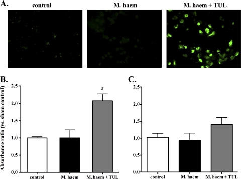
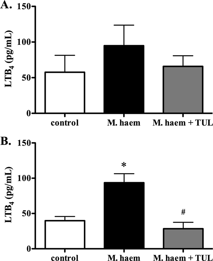
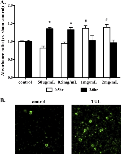
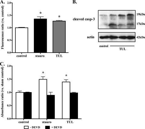
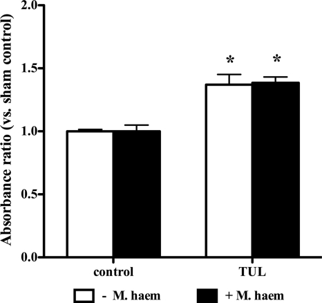
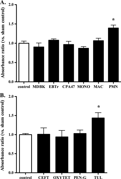
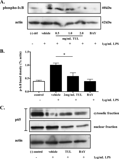
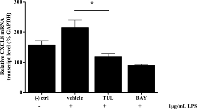
Clearance of apoptotic neutrophils is a central feature of the resolution of inflammation. Findings indicate that immuno-modulation and induction of neutrophil apoptosis by macrolide antibiotics generate anti-inflammatory benefits via mechanisms that remain obscure. Tulathromycin (TUL), a new antimicrobial agent for bovine respiratory disease, offers superior clinical efficacy for reasons not fully understood. The aim of this study was to identify the immuno-modulating effects of tulathromycin and, in this process, to establish tulathromycin as a new model for characterizing the novel anti-inflammatory properties of antibiotics. Bronchoalveolar lavage specimens were collected from Holstein calves 3 and 24 h postinfection, challenged intratracheally with live Mannheimia haemolytica (2 × 10(7) CFU), and treated with vehicle or tulathromycin (2.5 mg/kg body weight). Terminal deoxynucleotidyltransferase-mediated dUTP-biotin nick end labeling (TUNEL) staining and enzyme-linked immunosorbent assay (ELISA) revealed that tulathromycin treatment significantly increased leukocyte apoptosis and reduced levels of proinflammatory leukotriene B(4) in M. haemolytica-challenged calves. In vitro, tulathromycin concentration dependently induced apoptosis in freshly isolated bovine neutrophils from healthy steers in a capase-3-dependent manner but failed to induce apoptosis in bovine fibroblasts, epithelial cells, and endothelial cells, as well as freshly isolated bovine blood monocytes and monocyte-derived macrophages. The proapoptotic effects of TUL were also, in part, drug specific; equimolar concentrations of penicillin G, oxytetracycline, and ceftiofur failed to cause apoptosis in bovine neutrophils. In addition, tulathromycin significantly reduced levels of phosphorylated IκBα, nuclear translocation of NF-κB p65, and mRNA levels of proinflammatory interleukin-8 in lipopolysaccharide (LPS)-stimulated bovine neutrophils. The findings illustrate novel mechanisms through which tulathromycin confers anti-inflammatory benefits.


Figures

FIG. 1.
FIG. 1.
Tulathromycin induces cell apoptosis in calves challenged intratracheally with live M. haemolytica. (A) Fluorescent staining of bronchoalveolar lavage (BAL) samples fixed with 4% paraformaldehyde. Green represents TUNEL staining of apoptotic cells. Levels of apoptotic mono-/oligonucleosomes in BAL samples isolated from sham-treated (control), M. haemolytica-challenged calves (M. haem) and tulathromycin-treated calves challenged with M. haemolytica (M. haem + TUL) were measured using a cell death ELISA at 3 h (B) and 24 h (C) postinfection. Values were calculated as absorbance ratios versus values measured with cells from uninfected control calves. Values are means ± standard errors of the means. n = 5 to 8/group. *, P < 0.05.
FIG. 2.
FIG. 2.
Tulathromycin reduces levels of leukotriene B4 in calves challenged intratracheally with live M. haemolytica. LTB4 synthesis in the bronchoalveolar lavage fluid isolated from sham-treated calves (control), M. haemolytica-challenged calves (M. haem) and tulathromycin-treated calves challenged with M. haemolytica (M. haem + TUL) at 3 h (A) and 24 h (B) postinfection. Values are means ± standard errors of the means. n = 5 to 8/group. *, P < 0.05 versus control; #, P < 0.05 versus M. haemolytica-challenged calves.
FIG. 3.
FIG. 3.
Tulathromycin induces apoptosis in bovine neutrophils in a time- and concentration-dependent manner in vitro. (A) Levels of apoptotic mono-/oligonucleosomes in neutrophils incubated with tulathromycin (50 μg/ml to 2 mg/ml) for 0.5 h and 2.0 h were measured using a cell death ELISA. Values were calculated as absorbance ratios versus values measured in neutrophils from controls incubated with HBSS (control), arbitrarily set to 1.0. (B) Fluorescent staining of apoptotic neutrophils after 0.5 h of incubation with HBSS (control) or tulathromycin (TUL; 2 mg/ml), as determined by annexin V-FITC fluorescent staining of externalized phosphatidyl serine. Values are means ± standard errors of the means. n = 6/group. * and #, P < 0.05 versus control.
FIG. 4.
FIG. 4.
Induction of apoptosis in bovine neutrophils by tulathromycin is caspase-3 dependent. (A) Activity of caspase-3 in neutrophils treated with HBSS (control) or 2 mg/ml tulathromycin after 0.25 h. (B) Western blot analysis of cleaved caspase-3 in control and tulathromycin-treated (TUL; 2 mg/ml) bovine neutrophils for 0.25 h. Data illustrate 1 control and 3 tulathromycin-treated samples from 3 separate experiments. (C) Cell death ELISA of bovine neutrophils pretreated with a caspase-3 inhibitor (DEVD; 50 μM) for 1 h, followed by treatment with HBSS or tulathromycin (TUL; 2 mg/ml) for 0.5 h. Neutrophils treated with staurosporine (stauro; 1 μM) served as a positive control. Values are means ± standard errors of the means. n = 3 to 6/group. *, P < 0.05 versus control.
FIG. 5.
FIG. 5.
Tulathromycin induces apoptosis in bovine neutrophils in the presence and absence of live M. haemolytica. Levels of apoptotic mono-/oligonucleosomes in neutrophils incubated with HBSS (control) or tulathromycin (TUL; 2 mg/ml) for 0.5 h in the presence or absence of 2 × 10⁷ CFU M. haemolytica were measured using a cell death ELISA. Values were calculated as absorbance ratios versus values measured with neutrophils from controls incubated with HBSS, arbitrarily set to 1.0. Values are means ± standard errors of the means. n = 4 to 6/group. *, P < 0.05 versus control.
FIG. 6.
FIG. 6.
Tulathromycin-induced apoptosis is cell (A) and drug (B) selective. (A) Administration of tulathromycin increases levels of apoptotic mono-/oligonucleosomes in bovine neutrophils (PMN) but not in bovine epithelial cells (MDBK), fibroblasts (EBTr), endothelial cells (CPA47), freshly isolated blood monocytes (MONO), or monocyte-derived macrophages (MAC). (B) Tulathromycin (TUL; 2 mg/ml) but not ceftiofur (CEFT), oxytetracycline (OXYTET), or penicillin G (PEN-G) added at equimolar concentrations for 0.5 h increases levels of apoptosis in bovine neutrophils. Values were calculated as absorbance ratios versus values measured in neutrophils from control samples incubated with HBSS, arbitrarily set to 1.0. Values are means ± standard errors of the means. n = 4/group. *, P < 0.05 versus control.
FIG. 7.
FIG. 7.
Tulathromycin reduces phosphorylation of IκBα and prevents nuclear translocation of NF-κB p65. Representative immunoblotting (A) and densitometry analysis (B) of phosphorylated IκBα in LPS (1 μg/ml)-stimulated bovine neutrophils pretreated with MG132 proteosome inhibitor (20 μM), and cytosolic and nuclear p65 in LPS-stimulated bovine neutrophils treated with HBSS (vehicle) or tulathromycin (TUL; 2 mg/ml) for 0.5 h (C). BAY 11-7085 (BAY; 50 μM) inhibitor served as a positive control. Unstimulated neutrophils treated with HBSS served as a negative control. Densitometry data are expressed as percent actin. Values are means ± standard errors of the means. n = 3 to 4/group. *, P < 0.05 versus LPS-stimulated control.
FIG. 8.
FIG. 8.
Tulathromycin inhibits mRNA transcription of CXCL8. Real-time PCR measuring relative CXCL8 mRNA levels of bovine neutrophils coincubated with LPS (1 μg/ml) and HBSS (vehicle) or tulathromycin (TUL; 2 mg/ml) for 1.0 h. The inhibitor BAY 11-7085 (BAY; 50 μM) served as a positive control. Unstimulated neutrophils treated with HBSS served as a negative control. Values are means ± standard errors of the means. n = 4/group. *, P < 0.05 versus LPS-stimulated control.

Similar articles

Cited by

References

    1. Akgul, C., D. A. Moulding, and S. W. Edwards. 2001. Molecular control of neutrophil apoptosis. FEBS Lett. 487:318-322. - PubMed
    1. Aoki, Y., and P. N. Kao. 1999. Erythromycin inhibits transcriptional activation of NF-kappaB, but not NFAT, through calcineurin-independent signaling in T cells. Antimicrob. Agents Chemother. 43:2678-2684. - PMC - PubMed
    1. Aoshiba, K., A. Nagai, and K. Konno. 1995. Erythromycin shortens neutrophil survival by accelerating apoptosis. Antimicrob. Agents Chemother. 39:872-877. - PMC - PubMed
    1. Barkett, M., D. Xue, H. R. Horvitz, and T. D. Gilmore. 1997. Phosphorylation of IkappaB-alpha inhibits its cleavage by caspase CPP32 in vitro. J. Biol. Chem. 272:29419-29422. - PubMed
    1. Buret, A. G. 2010. Immuno-modulation and anti-inflammatory benefits of antibiotics: the example of tilmicosin. Can. J. Vet. Res. 74:1-10. - PMC - PubMed

Publication types

MeSH terms