

Epidemiology of ductal carcinoma in situ
- PMID: 20956818
- PMCID: PMC5161058
- DOI: 10.1093/jncimonographs/lgq027
Epidemiology of ductal carcinoma in situ
Abstract
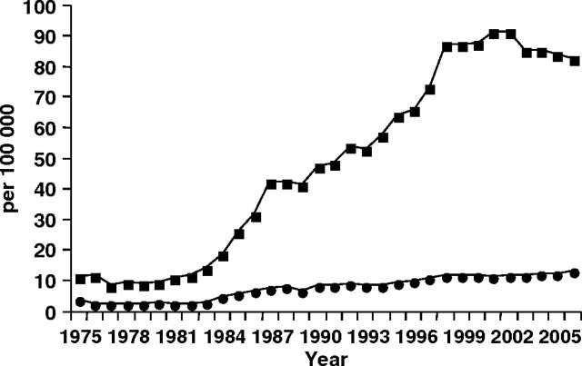
Ductal carcinoma in situ (DCIS) is a relatively common diagnosis among women undergoing screening mammography. The greatest increases in DCIS incidence have been in non-comedo subtypes of DCIS that are not associated with subsequent invasive cancer. After a 500% increase in DCIS from 1983 to 2003, the incidence of DCIS declined in women aged 50 years and older, whereas the incidence in women younger than age 50 continues to increase. Having undergone mammography is one of the strongest and most prevalent risk factors associated with a diagnosis of DCIS. Other risk factors for DCIS are similar to that for invasive cancer including increasing age, family history of breast cancer, high mammographic breast density, and postmenopausal hormone therapy use. Treatment for DCIS is relatively aggressive with the use of both surgery and radiation therapy and most recently adjuvant hormonal therapy. Breast cancer mortality is low and similar with all types of treatment. New information regarding incidence of DCIS and subtypes of DCIS according to frequency of mammography and risk factors could lead to insights into the biology of DCIS.
Figures
References
-
- White E, Lee CY, Kristal AR. Evaluation of the increase in breast cancer incidence in relation to mammography use. J Natl Cancer Inst. 1990;82(19):1546–1552. - PubMed
-
- Ernster V, Ballard-Barbash R, Barlow W, et al. Detection of DCIS in women undergoing screening mammography. J Natl Cancer Inst. 2002;94(20):1546–1554. - PubMed
-
- Kerlikowske K, Grady D, Barclay J, Sickles EA, Eaton A, Ernster V. Positive predictive value of screening mammography by age and family history of breast cancer. JAMA. 1993;270(20):2444–2450. - PubMed
-
- May D, Lee N, Richardson L, Giustozzi A, Bobo J. Mammography and breast cancer detection by race and Hispanic ethnicity: results from a national program (United States) Cancer Causes Control. 2000;11(8):697–705. - PubMed
-
- Jemal A, Siegel R, Ward E, Hao Y, Xu J, Thun M. Cancer statistics. CA Cancer J Clin. 2009. 2009;59(4):225–249. - PubMed
Publication types
MeSH terms
Grants and funding
LinkOut - more resources
Full Text Sources
Other Literature Sources
Medical
