. 2010 Dec 2;29(48):6402-8.
doi: 10.1038/onc.2010.360. Epub 2010 Oct 18.

c-Src differentially regulates the functions of microtentacles and invadopodia

Affiliations

c-Src differentially regulates the functions of microtentacles and invadopodia

E M Balzer et al. Oncogene. .

Abstract

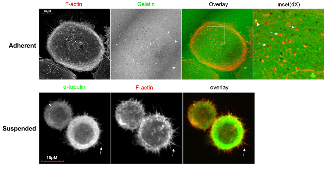
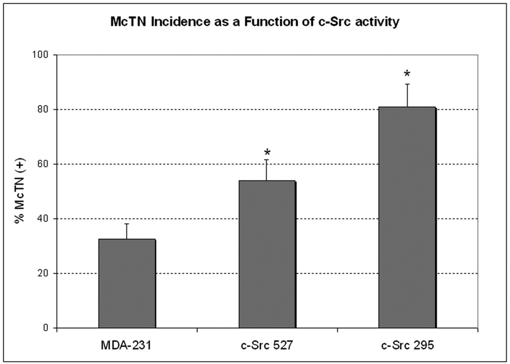
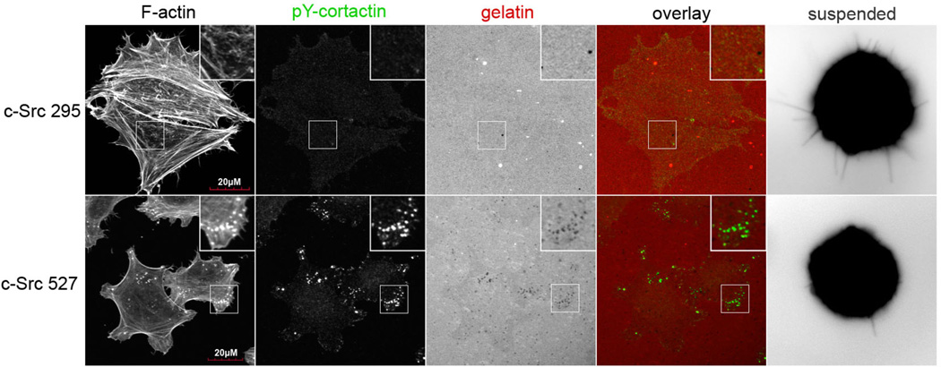
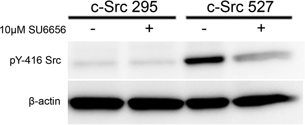
During metastasis, invading cells produce various actin-based membrane protrusions that promote directional migration and proteolysis of extracellular matrix (ECM). Observations of actin staining within thin, tubulin-based microtentacle (McTN) protrusions in suspended MDA-MB-231 tumor cells, prompted an investigation of whether McTNs are structural or functional analogs of invadopodia. We show here that MDA-MB-231 cells are capable of producing invadopodia and McTNs, both of which contain F-actin. Invadopodium formation was enhanced by the expression of a constitutively active c-Src kinase, and repressed by the expression of dominant-negative, catalytically inactive form of c-Src. In contrast, expression of inactive c-Src significantly increased McTN formation. Direct inhibition of c-Src with the SU6656 inhibitor compound also significantly enhanced McTN formation, but suppressed invadopodia, including the appearance of F-actin cores and phospho-cortactin foci, as well as completely blocking focal degradation of ECM. In addition, silencing of Tks5 in Src-transformed fibroblasts blocked invadopodia without affecting McTNs. Genetic modification of c-Src activity that promoted McTN formation augmented capillary retention of circulating tumor cells in vivo and rapid re-attachment of suspended cells in vitro, even though invadopodia were strongly suppressed. These results indicate that McTNs are capable of enhancing tumor cell reattachment, even in the absence of Tks5 and active Src, and define separate cytoskeletal mechanisms and functions for McTNs and invadopodia.


Conflict of interest statement

Conflict of interest. The authors declare no conflict of interest.

Figures

Figure 1
Figure 1. MDA-MB-231s form invadopodia and microtentacles, in a manner that is affected by c-Src activation
(A) Top row: When cultured for 4h on a thin layer of fluorescently-conjugated gelatin, MDA-MB-231 cells form focal areas of digested matrix that appear as dark spots or ‘holes’ (second panel) co-localizing with bright F-actin cores (Inset panel magnifies boxed region of overlay panel; arrowhead indicates an undigested region, while arrows point to degraded holes). Bottom row: Fixation of suspended cells revealed extensive plasma membrane protrusions (arrows) that co-stained for α-tubulin (left panel) and F-actin (middle panel). (B) McTN incidence is greatest in cells with dominant-negative c-Src295 (* denotes significant change relative to parental control, p<0.0001, n=12). (C) Immunofluorescence reveals that dominant-negative c-Src295 suppresses invadopodia formation and focal gelatin degradation, despite enhancing McTNs (suspended panel). Inset panels show 2X magnification of boxed region.
Figure 2
Figure 2. Acute c-Src inhibition inversely affects invadopodia and microtentacles
(A) Treatment of c-Src527 cells with SU6656 (10µM, 1h) suppressed phosphorylation of the activating tyrosine-416 residue, while c-Src295 cells showed only minimal kinase activation. (B) Exposure to 10µM SU6656 completely blocked invadopodia formation in c-Src-527 cells, as identified by co-labeling of phosphorylated cortactin puncta with F-actin cores, and gelatin holes. These features are clearly visible under vehicle control conditions (top row), but are lost when c-Src is inhibited (bottom row) Inset panels show 2X magnification of boxed region. (C) 10µM SU6656 enhanced the formation of McTNs in all cell lines examined. Arrows indicate long McTN extensions that resulted from c-Src inhibition (parental 231s, and c-Src-527s); comparably long extensions were observed basally in c-Src-295s (arrowheads), though at a lower frequency. (D) Blinded quantification revealed that McTN formation was significantly enhanced by SU6656, even in cells with low amounts of residual c-Src activity (* denotes significant changes relative to vehicle control, p<0.0001).
Figure 3
Figure 3. Tks5 silencing blocks invadopodia but does not affect McTN formation
(A) Src transformed NIH 3T3 cells (lane 2) were tested relative to clones that express non-silencing control shRNAs (C1 and C2: lanes 3 and 4), and shRNAs complementary to Tks5/Fish (4.20, and 4.24: lanes 5 and 6). These clones were lysed and total protein fractions were probed for Tks5/Fish via SDS-PAGE to demonstrate efficient Tks5 knockdown. Parental 3T3 cells are represented in lane 1, and an actin loading control is shown in the bottom row. (B) Gelatin degradation of Src 3T3 Tks5 clones was assayed by immunofluorescence. Src 3T3 and Src 3T3 C1 cells efficiently and rapidly formed invadopodia by 2 hours that co-localized with Tks5/Fish (inset panels, top and middle rows), while the Tks5 knockdown clone 4.24 showed a significant reduction in invadopodia formation and focal degradation, in agreement with previous reports [19]. (C) Microtentacle scoring over multiple independent trials (n=9) revealed an increase in McTNs among Src-transformed clones that was further enhanced by acute Src inhibition (t=30 minutes).
Figure 4
Figure 4. Capillary retention of circulating tumor cells is enhanced by alterations in c-Src activity
(A) Luciferase-expressing cells were injected into the tail vein of live mice and imaged in the presence of luciferin at selected timepoints, over a period of 2 days. c-Src295 cells were trapped and retained more efficiently in the lung capillaries throughout this time course, relative to MDA-231 parental cells. Reported are the average values of c-Src295 (n=14) and MDA-231 (n=10) over three independent trials. Asterisks denote significant differences in signal retention between cell lines (*: p<0.01, **: p=0.05). (B) Representative images were selected to demonstrate the retention over time of tumor cells in the lungs of a single mouse injected with either MDA-231 parental cells, or C-Src 295 cells. Temperature scale bar indicate absolute values for photon flux at each timepoint.

Similar articles

Cited by

References

    1. Yilmaz M, Christofori G. EMT, the cytoskeleton, and cancer cell invasion. Cancer Metastasis Rev. 2009;28(1–2):15–33. - PubMed
    1. Gupton SL, Gertler FB. Filopodia: the fingers that do the walking. Sci STKE. 2007;2007(400):re5. - PubMed
    1. Machesky LM. Lamellipodia and filopodia in metastasis and invasion. FEBS Lett. 2008;582(14):2102–2111. - PubMed
    1. Linder S. The matrix corroded: podosomes and invadopodia in extracellular matrix degradation. Trends Cell Biol. 2007;17(3):107–117. - PubMed
    1. Buccione R, Caldieri G, Ayala I. Invadopodia: specialized tumor cell structures for the focal degradation of the extracellular matrix. Cancer Metastasis Rev. 2009;28(1–2):137–149. - PubMed

Publication types