. 2010 Oct 1;5(10):e13094.
doi: 10.1371/journal.pone.0013094.

Monomerization of cytosolic mature smac attenuates interaction with IAPs and potentiation of caspase activation

Affiliations

Monomerization of cytosolic mature smac attenuates interaction with IAPs and potentiation of caspase activation

Stephen P Burke et al. PLoS One. .

Abstract

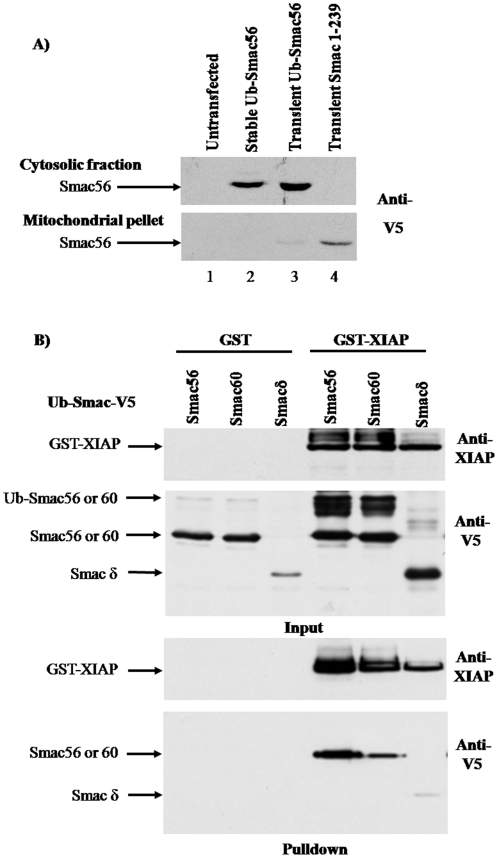
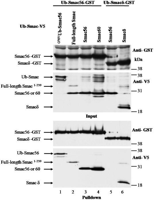
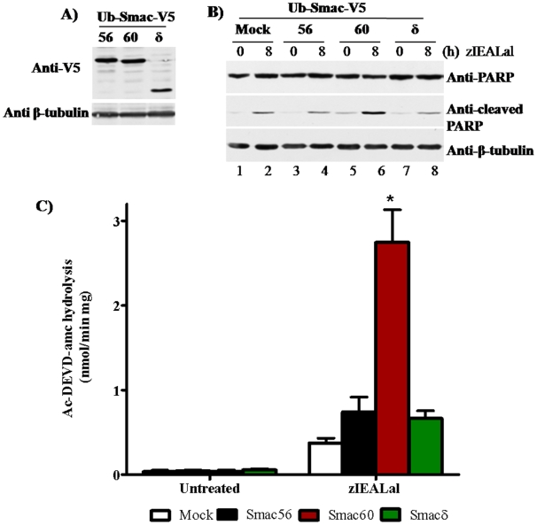
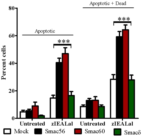
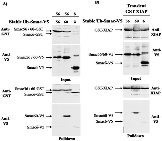
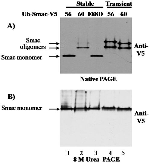
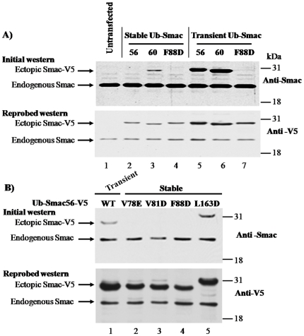
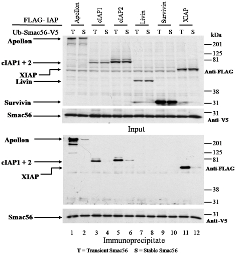
The four residues at the amino-terminus of mature Smac/DIABLO are an IAP binding motif (IBM). Upon exit from mitochondria, mature Smac interacts with inhibitor of apoptosis proteins (IAPs), abrogating caspase inhibition. We used the ubiquitin fusion model to express mature Smac in the cytosol. Transiently expressed mature Smac56-239 (called Smac56) and Smac60-239 (called Smac60), which lacks the IBM, interacted with X-linked inhibitor of apoptosis protein (XIAP). However, stable expression produced wild type Smac56 that failed to homodimerize, interact with XIAP, and potentiate caspase activation. Cytosolic Smac60 retained these functions. Cytosolic Smac56 apparently becomes posttranslationally modified at the dimer interface region, which obliterated the epitope for a monoclonal antibody. Cytosolic Smacδ, which has the IBM but lacks amino acids 62-105, homodimerized and weakly interacted with XIAP, but failed to potentiate apoptosis. These findings suggest that the IBM of Smac is a recognition point for a posttranslational modification(s) that blocks homodimerization and IAP interaction, and that amino acids 62-105 are required for the proapoptotic function of Smac.


Conflict of interest statement

Competing Interests: The authors have declared that no competing interests exist.

Figures

Figure 1
Figure 1. Cytosolic expression of mature Smac variants and the interaction of Smac56, Smac60, and Smacδ with XIAP following transient expression.
(A) Cells were stably or transiently transfected with IRES2.eGFP expression plasmid (Stable versus Transient) containing Ub-Smac56 or full-length Smac 1-239. 911 cells were subjected to hypotonic lysis, and cytosol and mitochondrial fractions were prepared by differential centrifugation. Proteins were resolved by SDS-PAGE and subjected to western blot analysis with a V5 antibody to detect ectopic Smac. (B) Cells were transiently cotransfected with the indicated GST vector (pGFLEX) and the indicated HA-Ub-Smac (pcDNA3.1-V5His). Two days later cells were lysed, and proteins were purified using GSH-agarose. Lysate proteins (Input) and proteins eluted from the GSH-agarose (Pulldown) were resolved by SDS-PAGE and subjected to western blot analysis with antibody to the V5 epitope or XIAP.
Figure 2
Figure 2. Transiently expressed Smac56 homodimerizes and heterodimerizes with Smac60, but not with Smacδ.
Cells were transiently cotransfected with the Ub-Smac56 or Ub-Smacδ with a C-terminal GST tag (pGFLEX) and with full-length Smac1-239, Ub-Smac56, Ub-Smac60, Ub-Smacδ, or G76VUb-Smac56 with a C-terminal V5 tag (pcDNA3.1-HA-Ub-Smac-V5), as indicated. Cells were lysed, and proteins were purified using GSH-agarose. Lysate proteins (Input) and proteins eluted from the GSH-agarose (Pulldown) were resolved by SDS-PAGE and subjected to western blot analysis with antibody to GST-pi and to the V5 tag.
Figure 3
Figure 3. Persistent cytosolic expression of Smac60, but not Smac56, potentiates activation of caspase-3-like activity by zIEALal.
(A) Cells, which stably expressed the indicated Smac variant in the IRES2.eGFP vector, were lysed and proteins (30 µg) were subjected to western blot analysis with V5 antibody. (B) Stable Smac cell lines (IRES2.eGFP) were plated, and the following day they were treated for 8 h with 20 µM zIEALal or left untreated. Cell lysates were subjected to western blot analysis for intact PARP (B, top panel) or PARP cleaved after aspartate 214 (B, middle panel). Anti β-tubulin western blot confirmed equivalent protein loading (B, bottom panel). (C) Cell lines were treated for 18 h with 20 µM zIEALal or untreated. Lysates (0.75 µg) were assayed for caspase-3-like activity by continuously recording fluorescence of amc produced by hydrolysis of Ac-DEVD-amc. Columns indicate mean amc produced ± S.E.M. Two-way Anova followed by the Bonferroni post-test gave a p value of <0.001 for zIEALal treated Smac60 compared to the empty IRES vector (n = 4) (Prism 5, Graphpad Software). Mock indicates cell lines stably transfected with empty vector.
Figure 4
Figure 4. Persistent cytosolic expression of Smac56 or Smac60 potentiates apoptosis and cell death evoked by zIEALal.
Cell lines that stably expressed the indicated Smac variant (3 independent clones transfected with empty vector (IRES2.eGFP) (Mock) or with the indicated Smac variant) were incubated with or without zIEALal for 24 h. Following the treatment, both floating and attached cells were harvested and stained with annexin V-PE and 7-AAD, and analyzed by FACS. Apoptotic cells positively stained for annexin V-PE only, and dying cells were positively stained for 7-AAD only or for 7-AAD and annexin V-PE. Values are mean ± S.E.M (n = 9). Two-way Anova followed by the Bonferroni post-test gave the indicated p values (***<0.001) (Prism, Graphpad Software).
Figure 5
Figure 5. Persistent cytosolic expression of Smac56, but not Smac60, ablates dimerization and interaction with XIAP.
(A) Cells that stably expressed the indicated Ub-Smac-V5 plasmid (IRES2.eGFP) were transiently transfected with the Ub-Smac-GST plasmids (pGFLEX), lysed, and proteins were purified using GSH-agarose. Lysate proteins (Input) and proteins eluted from the GSH-agarose (Pulldown) were resolved by SDS-PAGE and subjected to western blot analysis with antibody to GST-pi and the V5 epitope. (B) Cells that stably expressed the indicated Ub-Smac-V5 plasmid (IRES2.eGFP) were transiently transfected with GST-XIAP plasmid (pGFLEX), lysed, and Input and Pulldown proteins were resolved by SDS-PAGE and subjected to western blot analysis with antibody to XIAP and the V5 epitope as described for (A).
Figure 6
Figure 6. Native and urea PAGE of Smac56, Smac60, and the dimer interface F88D mutant of Smac56.
Cell lines that stably expressed the indicated Ub-Smac-V5 (IRES2.eGFP) and cells that were transiently transfected with Ub-Smac56-V5 or Ub-Smac60-V5 (IRES2.eGFP) were lysed, and proteins were resolved by native PAGE or by PAGE in the presence of 8 M urea as indicated. Western analysis was done with antibody to the V5 epitope.
Figure 7
Figure 7. Persistent cytosolic expression of Smac56, but not Smac60, ablates immunostaining by a monoclonal antibody similarly to certain missense mutations of the dimer interface.
(A) Cell lines that stably expressed the indicated Ub-Smac-V5 plasmids (IRES2.eGFP) and cells that were transiently transfected with Ub-Smac56-V5 or Ub-Smac60-V5 (IRES2.eGFP) were lysed and proteins were resolved by SDS-PAGE. Mock indicates a cell line transfected with empty IRES2.eGFP vector. Western blot analysis was done firstly with the monoclonal Smac antibody and secondly with antibody to the V5 epitope. (B) Cell lines that stably expressed the indicated missense mutation of Ub-Smac56-V5 (IRES2.eGFP) and cells that were transiently transfected with Ub-Smac56-V5 (IRES2.eGFP) were lysed, and proteins were resolved by SDS-PAGE. Western blot analysis was done as indicated for (A).
Figure 8
Figure 8. Persistent cytosolic expression of Smac56 attenuates its interaction with Apollon and cIAPs.
Cells indicated by a T (Transient) were cotransfected with Ub-Smac56-V5 (IRES2.eGFP), whereas those indicated by a S (Stable) stably expressed Ub-Smac56-V5 (IRES2.eGFP). Additional cultures were transiently transfected with the indicated FLAG-IAP (p3XFLAG.CMV10). The cells that stably expressed Ub-Smac56-V5 were treated for 12 h with 20 µM zIEALal to increase the level of Smac56, which is rapidly degraded by UPS (Burke and Smith, unpublished data). Cell pellets from each IAP transfection were combined with a pellet of cells that were transiently or stably transfected with Ub-Smac56. The combined cell pellets were lysed, and proteins were immunoprecipitated with a polyclonal antibody to the V5 epitope. Lysate proteins (Input) and proteins eluted from the immune complex (Immunoprecipitate) were resolved by SDS-PAGE and subjected to western blot analysis with monoclonal antibody to the V5 or FLAG epitope.

Similar articles

Cited by

References

    1. Du C, Fang M, Li Y, Li L, Wang X. Smac, a mitochondrial protein that promotes cytochrome c-dependent caspase activation by eliminating IAP inhibition. Cell. 2000;102:33–42. - PubMed
    1. Verhagen AM, Ekert PG, Pakusch M, Silke J, Connolly LM, et al. Identification of DIABLO, a mammalian protein that promotes apoptosis by binding to and antagonizing IAP proteins. Cell. 2000;102:43–53. - PubMed
    1. Roberts DL, Merrison W, MacFarlane M, Cohen GM. The inhibitor of apoptosis protein-binding domain of Smac is not essential for its proapoptotic activity. J Cell Biol. 2001;153:221–228. - PMC - PubMed
    1. Fu J, Jin Y, Arend LJ. Smac3, a novel Smac/DIABLO splicing variant, attenuates the stability and apoptosis-inhibiting activity of X-linked inhibitor of apoptosis protein. J Biol Chem. 2003;278:52660–52672. - PubMed
    1. Burri L, Strahm Y, Hawkins CJ, Gentle IE, Puryer MA, et al. Mature DIABLO/Smac is produced by the IMP protease complex on the mitochondrial inner membrane. Mol Biol Cell. 2005;16:2926–2933. - PMC - PubMed

Publication types

MeSH terms

Substances