. 2010 Oct 1;5(10):e13108.
doi: 10.1371/journal.pone.0013108.

PHOX2B-mediated regulation of ALK expression: in vitro identification of a functional relationship between two genes involved in neuroblastoma

Affiliations

PHOX2B-mediated regulation of ALK expression: in vitro identification of a functional relationship between two genes involved in neuroblastoma

Tiziana Bachetti et al. PLoS One. .

Abstract

Background: Neuroblastoma (NB) is a severe pediatric tumor originating from neural crest derivatives and accounting for 15% of childhood cancer mortality. The heterogeneous and complex genetic etiology has been confirmed with the identification of mutations in two genes, encoding for the receptor tyrosine kinase Anaplastic Lymphoma Kinase (ALK) and the transcription factor Paired-like Homeobox 2B (PHOX2B), in a limited proportion of NB patients. Interestingly, these two genes are overexpressed in the great majority of primary NB samples and cell lines. These observations led us to test the hypothesis of a regulatory or functional relationship between ALK and PHOX2B underlying NB pathogenesis.

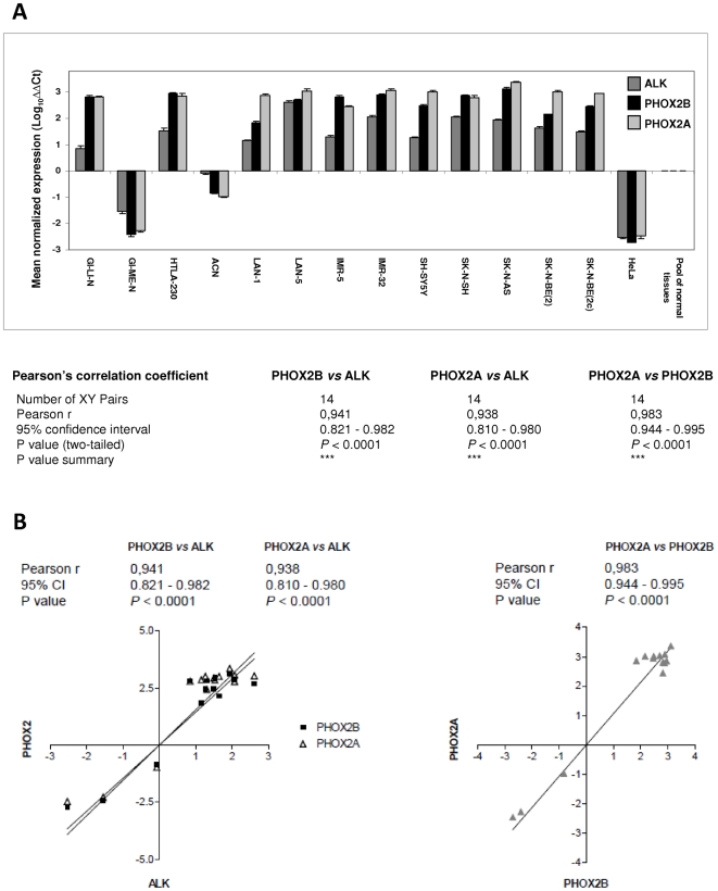
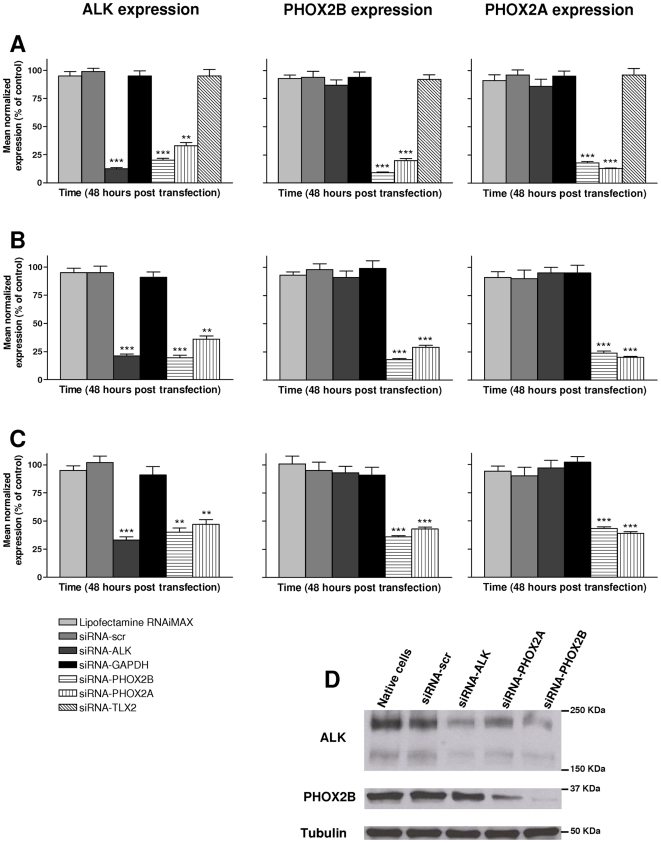
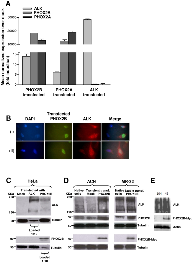
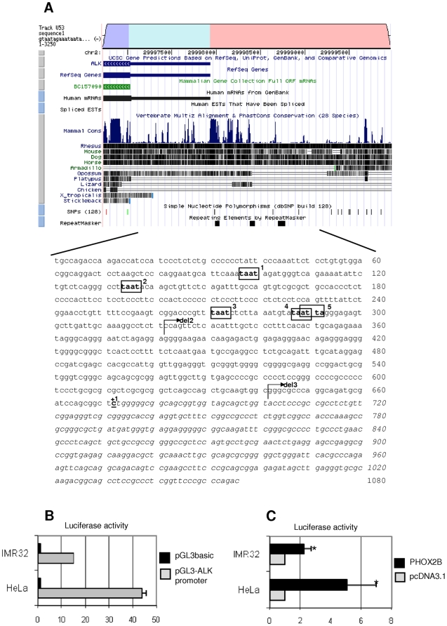
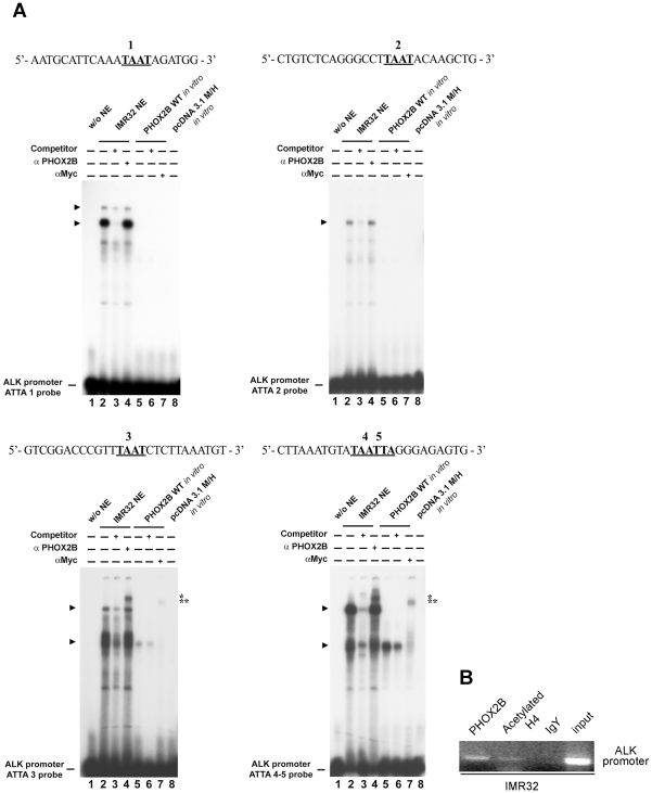
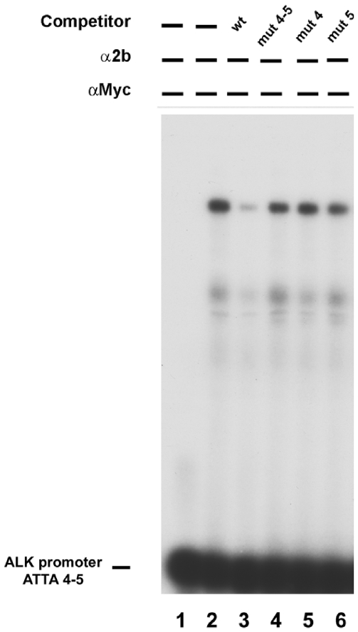
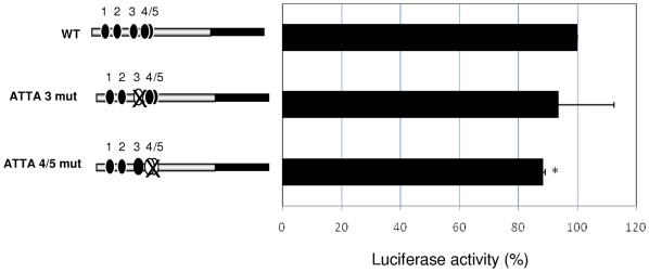
Methodology/principal findings: Following this possibility, we first confirmed a striking correlation between the transcription levels of ALK, PHOX2B and its direct target PHOX2A in a panel of NB cell lines. Then, we manipulated their expression in NB cell lines by siRNA-mediated knock-down and forced over-expression of each gene under analysis. Surprisingly, PHOX2B- and PHOX2A-directed siRNAs efficiently downregulated each other as well as ALK gene and, consistently, the enhanced expression of PHOX2B in NB cells yielded an increment of ALK protein. We finally demonstrated that PHOX2B drives ALK gene transcription by directly binding its promoter, which therefore represents a novel PHOX2B target.

Conclusions/significance: These findings provide a compelling explanation of the concurrent involvement of these two genes in NB pathogenesis and are going to foster a better understanding of molecular interactions at the base of the disease. Moreover, this work opens new perspectives for NBs refractory to conventional therapies that may benefit from the design of novel therapeutic RNAi-based approaches for multiple gene targets.


Conflict of interest statement

Competing Interests: The authors have declared that no competing interests exist.

Figures

Figure 1
Figure 1. Gene expression analysis in NB and HeLa cells and correlation analysis.
(A) Relative gene expression analysis of the ALK, PHOX2B and PHOX2A genes, carried out in a panel of NB and in HeLa cell lines by real-time RT-qPCR using a pool of normal tissue RNAs as reference sample (see Methods), shows over-expression of the three genes in all but two NB cell lines tested (GI-ME-N and ACN). (B) X-Y Plots showing a significant correlation between the expression level of PHOX2B and PHOX2A vs.ALK (left) and PHOX2Avs. PHOX2B (right) genes in the analyzed cell lines. Pearson's correlation coefficient indicates a very significant correlation of the three transcription levels vs. each other. Values are the mean ± s.d. of N = 3 independent RT-qPCR analyses performed in triplicate.
Figure 2
Figure 2. siRNA-mediated silencing of ALK, PHOX2B and PHOX2A in NB cells.
Effects on the transcription level of the ALK (left side graphs), PHOX2B (middle graphs) and PHOX2A (right side graphs) genes after knock-down of the same genes in SHSY-5Y (A), IMR-32 (B) and HTLA-230 (C) cells. Gene-specific knock-down, evaluated 48 hours post-transfection by real-time RT-qPCR analysis, is very effective but also PHOX2-directed siRNAs are able to downregulate ALK at a similar extent (**: P<0.01; ***: P<0.001). Values are the mean ± s.d. of N = 3 independent experiments performed in duplicate. (D) Gene silencing was confirmed at 72 hours post-transfection by Western blot.
Figure 3
Figure 3. Forced over-expression of ALK, PHOX2B and PHOX2A in HeLa and NB cell lines.
A) Transcription levels of the ALK, PHOX2B and PHOX2A genes were evaluated in HeLa cells 48 hours post-transfection with the corresponding gene-specific cDNA expressing vectors by real-time RT-qPCR. Besides the dramatic increase of gene transcripts by each respective transfectants, ALK expression results enhanced by the PHOX2 genes over-expression. Values are the mean ± s.d. of N = 3 independent experiments performed in duplicate. B) Upper (I) and lower (II) lanes from immunofluorescence analysis report examples of HeLa cells transfected with the PHOX2B-Myc expression construct. From left to right images show DAPI stained cell nuclei (blue), staining for the PHOX2B-Myc protein (green), and staining for the ALK protein (red). The most distal image is the merge of the three nearby figures. C) Western blot evaluating protein amounts of ALK and PHOX2B in HeLa cells at 72 hours post-transfection with gene-specific cDNA constructs. A consistent transcript increment of each gene is observed but ALK starts to be expressed also following the forced expression of PHOX2B-Myc protein. Gel was loaded with 100 µg of total protein extracts except for evaluation of ALK in ALK-transfected cells and PHOX2B in PHOX2B-transfected cells, for which 1∶10 (10 µg) of protein extracts was loaded to avoid over-saturation of autoradiograph films. D) Western blot evaluating protein amounts of ALK in ACN cells (left) at 72 hours post-transfection with the PHOX2B-Myc construct, in a clone of IMR-32 cells stably expressing the same PHOX2B-Myc fusion protein (right). A marked increment in ALK expression is detected following PHOX2B-Myc expression in transient –transfected ACN cells compared to both native and mock-transfected cells and in a clone of IMR-32 cells, stably expressing PHOX2B-Myc, compared to native cells. An anti-cMyc antibody was specifically used to distinguish PHOX2B-Myc fusion protein from endogenous PHOX2B of NB cells (lower blots). E) Two stable IMR-32 clones, one negative (104) and one positive (49) for PHOX2B-Myc expression were analyzed for ALK expression (upper panels); expression of the fusion PHOX2B-Myc protein was assessed by Western Blot using anti –cMyc (middle panels) and the anti-actin antibodies (bottom panels).
Figure 4
Figure 4. In silico representation of the subcloned ALK regulatory region.
A) In the upper section of the figure, a portion of the ALK gene, as shown by the Vista Browser, is reported: the ALK coding region (dark grey), the predicted 5′ untranslated region (light grey), part of the 5′ regulatory region (black) and their levels of phylogenetic conservation (dark bars) are represented. In the lower part, the sequence corresponds to the part of the gene subcloned into the pGL3basic vector, containing 672 bp upstream the putative transcriptional start site (indicated with +1) and 384 bp of the 5′ untranslated region (shown in Italic). Moreover, the five ATTA sites are boxed and progressively named from 1 to 5. Arrows represent the start point of the deletion constructs. B) Activity of the pGL3basic-ALK promoter construct (grey bars) in HeLa and IMR-32 cell lines, expressed as fold induction with respect to the empty pGL3basic vector (black bars). Values are the mean ± s.d. of N = 3 independent experiments performed in duplicate. C) ALK promoter induction in HeLa and IMR-32 cells co-transfected with the PHOX2B expression plasmid and expressed as fold induction of the Luciferase activity with respect to cells transfected with the empty vector (pcDNA3.1, arbitrary value = 1) (*: P<0.05).
Figure 5
Figure 5. In vitro interaction of PHOX2B with the ALK promoter.
A) EMSAs were performed using probes containing one of the ATTA sites of the region under analysis (ATTA 1, ATTA 2, ATTA 3 and the complex ATTA 4/5). Each labeled probe was incubated in the absence of nuclear extracts (lane 1), with IMR-32 nuclear extracts (lanes 2–4) or the in vitro expressed PHOX2B-Myc fusion protein (lanes 5–7). As negative control the oligonucleotides were also incubated with the in vitro reaction performed using the empty vector pcDNA3.1 M/H (lane 8). The competition experiments were performed in the presence of a molar excess of the unlabeled oligonucleotides (lanes 3 and 6). The anti-PHOX2B or the anti-c-Myc antibodies were added to the samples run in lanes 4 and 7, respectively. On the left, the arrows indicate the specific retarded bands detected; on the right, one or two asterisks indicate the supershifted complexes containing PHOX2B obtained by incubation of IMR-32 nuclear extracts with the anti-PHOX2B antibody (*) or the in vitro expressed protein with the anti-cMyc antibody (**), respectively. The free probes are shown at the bottom of the gels. B) ChIP assay. Chromatin extracted from IMR-32 cells was immunoprecipitated using the antibody against PHOX2B; pre-immune chicken IgY and the anti-acetylated histone H4 antibodies were used as negative and positive controls, respectively. The input represent 0,5% of the total chromatin extract. The precipitated DNA has undergone PCR amplification by using primers bordering the ATTA 3 and the ATTA 4/5 boxes in the ALK promoter.
Figure 6
Figure 6. Effect of ATTA 4/5 disruption in competition of PHOX2B binding.
IMR32 nuclear extracts were incubated with the ATTA4/5 probe (lane 2) and competition obtained by adding an excess of: a wild type (wt) probe (lane 3), a probe mutated in both ATTA 4 and ATTA 5 sites (lane 4) or in each of them (lanes 5–6). Incubation without nuclear extracts was regarded as negative control (lane 1).
Figure 7
Figure 7. Effects of mutagenesis of ATTA 3 and ATTA 4/5 on the PHOX2B-mediated ALK trans-activation.
Left side: schematic representation of the three constructs carrying all the ATTA boxes functional (wt, all four black circles), the ATTA 3 disrupted (ATTA 3 mut, one white circle) or both the ATTA 4 and 5 disrupted (ATTA 4/5 mut, two white circles). Right side: induction of the ALK promoter containing the mutant ATTA 3 and ATTA 4/5 in HeLa cells co-transfected with the PHOX2B expression plasmid are expressed as percentage of the Luciferase activity obtained by cells co-transfected with PHOX2B and the ALK promoter (wt) vectors (wt, arbitrary value = 100). Values are the mean ± s.d. of N = 3 independent experiments (*: P<0.05).
Figure 8
Figure 8. PHOX2B effect on the ALK promoter sequentially deleted plasmids.
A) Schematic representation of deleted plasmid inserts, progressively shorter from the entire wt ALK promoter region considered (−671 bp), down to the so called deletion 2 (del2; −351 bp), and to the so called deletion 3 (del3; −31 bp) (see also figure 4A). The promoter (grey bar), the 5′UTR (black bar) and the ATTA boxes (black circles) are shown. B) Activity of the ALK promoter fragments, expressed as percentage of the activity of the wt construct. Values are the mean ± s.d. of N = 3 independent experiments performed in HeLa cells. C) PHOX2B-mediated induction of the ALK promoter deleted plasmids, expressed as fold increase of the Luciferase activity obtained with respect to the use of the empty vector (pcDNA3.1) on the wt promoter. Values are the mean ± s.d. of N = 3 independent experiments performed in duplicate in HeLa cells.

Similar articles

Cited by

References

    1. Trochet D, Bourdeaut F, Janoueix-Lerosey I, Deville A, de Pontual L, et al. Germline mutations of the paired-like homeobox 2B (PHOX2B) gene in neuroblastoma. Am J Hum Genet. 2004;74:761–764. - PMC - PubMed
    1. Trochet D, O'Brien LM, Gozal D, Trang H, Nordenskjold A, et al. PHOX2B genotype allows for prediction of tumor risk in congenital central hypoventilation syndrome. Am J Hum Genet. 2005;76:421–426. - PMC - PubMed
    1. Mosse YP, Laudenslager M, Longo L, Cole KA, Wood A, et al. Identification of ALK as a major familial neuroblastoma predisposition gene. Nature. 2008;455:930–935. - PMC - PubMed
    1. Pattyn A, Morin X, Cremer H, Goridis C, Brunet JF. The homeobox gene Phox2b is essential for the development of autonomic neural crest derivatives. Nature. 1999;399:366–370. - PubMed
    1. Adachi M, Browne D, Lewis EJ. Paired-like homeodomain proteins Phox2a/Arix and Phox2b/NBPhox have similar genetic organization and independently regulate dopamine beta-hydroxylase gene transcription. DNA Cell Biol. 2000;19:539–554. - PubMed

Publication types

MeSH terms