. 2010 Oct 1;5(10):e13109.
doi: 10.1371/journal.pone.0013109.

Failure of effector function of human CD8+ T Cells in NOD/SCID/JAK3⁻/⁻ immunodeficient mice transplanted with human CD34+ hematopoietic stem cells

Affiliations

Failure of effector function of human CD8+ T Cells in NOD/SCID/JAK3⁻/⁻ immunodeficient mice transplanted with human CD34+ hematopoietic stem cells

Yoshinori Sato et al. PLoS One. .

Abstract

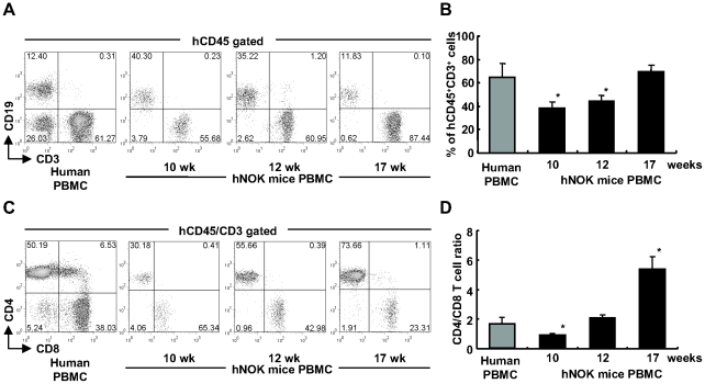
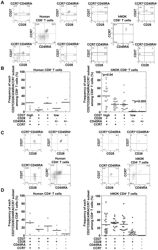
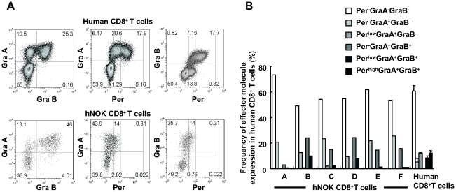
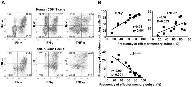
Humanized mice, which are generated by transplanting human CD34+ hematopoietic stem cells into immunodeficient mice, are expected to be useful for the research on human immune responses. It is reported that antigen-specific T cell responses occur in immunodeficient mice transplanted with both human fetal thymus/liver tissues and CD34+ fetal cells, but it remains unclear whether antigen-specific T cell responses occur in those transplanted with only human CD34+ hematopoietic stem cells (HSCs). Here we investigated the differentiation and function of human CD8+ T cells reconstituted in NOD/SCID/Jak3⁻/⁻ mice transplanted with human CD34+ HSCs (hNOK mice). Multicolor flow cytometric analysis demonstrated that human CD8+ T cells generated from the CD34+ HSCs comprised only 3 subtypes, i.e., CD27(high)CD28+CD45RA+CCR7+, CD27+CD28+CD45RA⁻CCR7+, and CD27+CD28+CD45RA⁻CCR7⁻and had 3 phenotypes for 3 lytic molecules, i.e., perforin(Per)⁻granzymeA(GraA)⁻granzymeB(GraB)⁻, Per⁻GraA+GraB⁻, and Per(low)GraA+GraB+. These CD8+ T cells failed to produce IFN-γ and to proliferate after stimulation with alloantigens. These results indicate that the antigen-specific T cell response cannot be elicited in mice transplanted with only human CD34+ HSCs, because the T cells fail to develop normally in such mice.


Conflict of interest statement

Competing Interests: The authors have declared that no competing interests exist.

Figures

Figure 1
Figure 1. Flow cytometric analysis of human T cells reconstituted in hNOK mice.
(A) A representative result for hCD45+CD3+ T cells and hCD45+CD19+ B cells among PBMC from hNOK mice at 10, 12, and 17 weeks after the transplantation. (B) Summarized results showing hCD45+CD3+ T cell proportion in PBMC from hNOK mice (n = 20) at 10, 12 and 17 weeks. (C) A representative result for human CD4+ and CD8+ T cell proportions in PBMC from hNOK at 10, 12 and 17 weeks after the transplantation. (D) Summarized results showing human CD4/CD8 T cell ratio for PBMC from hNOK mice at 10, 12, and 17 weeks. Human PBMC from healthy adult individuals (n = 4) are shown as a control. Bar graph data are shown as the mean ± SEM of 7 independent experiments. *, p<0.05, human PBMC vs. hNOK mouse PBMC.
Figure 2
Figure 2. Phenotypic classification of human CD8+ T cells reconstituted in hNOK mice.
Human T cells among PBMC from hNOK mice at 17 weeks after the transplantation were analyzed for the expression of the following cell-surface markers: CD4, CD8, CD27, CD28, CD45RA, and CCR7. (A) A representative result of 5-color flow cytometric analysis of the human CD8+ T cell population among PBMC from a hNOK mouse (right data) and an adult human (left data). (B) Summarized results showing frequency of subsets among the human CD8+ T cells isolated from the PBMC from hNOK mice (n = 20, right data) and human adult individuals (n = 4, left data). (C) A representative result of 5-color flow cytometric analysis of human CD4+ T cell population in PBMC from hNOK mice (right data) and a human adult individual (left data). (D) Summarized result showing human CD4+ T cell proportion in PBMC from hNOK mice (n = 20, right data) and human adult individuals (n = 4, left data). Each symbol represents 1 mouse; the mean value is shown as a horizontal solid line. *, p<0.05; **, p<0.01, human PBMC vs. hNOK mice PBMC.
Figure 3
Figure 3. Expression of 3 cytolytic effector molecules of human CD8+ T cells reconstituted in hNOK mice.
Human CD8+ T cells among splenocytes of hNOK mice were analyzed for the expression of 3 cytolytic effector molecules: Per, Gra A, and Gra B. (A) A representative result showing co-expression of 3 cytolytic effector molecules in human CD8+ T cells from a hNOK mouse (lower data) and a human adult individual (upper data). (B) Summarized result for the expression of 3 the cytolytic effector molecules in human CD8+ T cells from hNOK mice (n = 6) and human adult individuals (n = 3).
Figure 4
Figure 4. Cytokine production of human CD8+ T cells reconstituted in hNOK mice.
Splenocytes from hNOK mice were cultured with PMA and ionomycin for 6 hours and then the production of IFN-γ, TNF-α, and IL-2 cytokines by human CD8+ T cells was measured by flow cytometry. (A) A representative result for cytokine production by human CD8+ T cells from a hNOK mouse (lower data) and a human adult individual (upper data). (B) Correlation between frequency of IFN-γ-, TNF-α-, and IL-2+IFN-γTNF-α (IL-2single+)-producing CD8+ T cells and that of effector memory (CD27+CD28+CCR7CD45RA plus CD27lowCD28CD45RA+/−CCR7) CD8+ T cell subset (n = 12).
Figure 5
Figure 5. Alloreactivity of human CD8+ T cells reconstituted in hNOK mice.
The hNOK mice were immunized for 31 days with irradiated human PBMC (allo-hPBMC) from a healthy donor with HLA-A*2402/A*2402, HLA-B*5201/B*5901, and HLA-DRB1*1502/DRB1*0405 (immunized mice) or PBS (un-immunized mice). The splenocytes of each hNOK mice were harvested, and the cells were stained by using anti-human CD45, anti-human CD3, and anti-human CD8 mAbs. (A) Human adult PBMC from a healthy donor with HLA-A*2601/A*2403, HLA-B*3501/B*5101, and HLA-DRB1*0405/DRB1*0405 were cultured with the allo-hPBMC or the self-hPBMC for 7 days in vitro, and a representative result showing the IFN-γ production by the human CD8+ T cells and the human CD8 T cells is shown as a control (upper data). The splenocytes from the immunized hNOK mice were cultured with the allo-hPBMC or self-CB for 7 days in vitro, and a representative result for IFN-γ production by the human CD8+ T cells and the human CD8 T cells is shown (lower data). (B) Splenocytes from the immunized hNOK mice were cultured with irradiated 721.221 cells expressing HLA-A*2402 (.221-A*2402) or irradiated 721.221 expressing HLA-B*5201 (.221-B*5201) for 3 days in vitro, and a representative result for the proliferation of the human CD8+ T cells or the human CD8 T cells is shown. One representative experiment from 3 experiments is shown.

Similar articles

Cited by

References

    1. Shultz LD, Ishikawa F, Greiner DL. Humanized mice in translational biomedical research. Nat Rev Immunol. 2007;7:118–30. - PubMed
    1. Manz MG. Human-hemato-lymphoid-system mice: opportunities and challenges. Immunity. 2007;26:537–541. - PubMed
    1. Legrand N, Weijer K, Spits H. Experimental models to study development and function of the human immune system in vivo. J Immunol. 2006;176:2053–2058. - PubMed
    1. Hiramatsu H, Nishikomori R, Heike T, Ito M, Kobayashi K, et al. Complete reconstitution of human lymphocytes from cord blood CD34+ cells using the NOD/SCID/γc null mice model. Blood. 2003;102:873–880. - PubMed
    1. Ishikawa F, Yasukawa M, Lyons B, Yoshida S, Miyamoto T, et al. Development of functional human blood and immune systems in NOD/SCID/IL2 receptor γ chainnull mice. Blood. 2005;106:1565–1573. - PMC - PubMed

Publication types