. 2010 Oct 1;5(10):e13134.
doi: 10.1371/journal.pone.0013134.

Casein kinase 2 dependent phosphorylation of neprilysin regulates receptor tyrosine kinase signaling to Akt

Affiliations

Casein kinase 2 dependent phosphorylation of neprilysin regulates receptor tyrosine kinase signaling to Akt

Martin Siepmann et al. PLoS One. .

Abstract

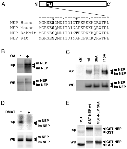
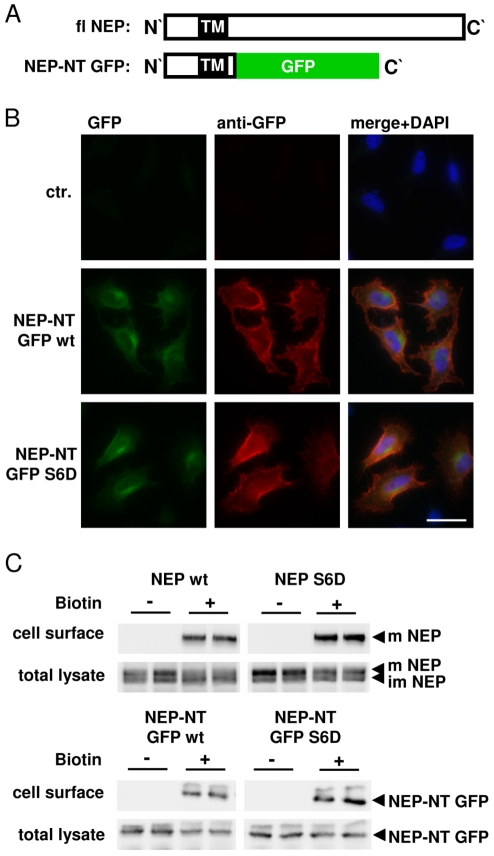
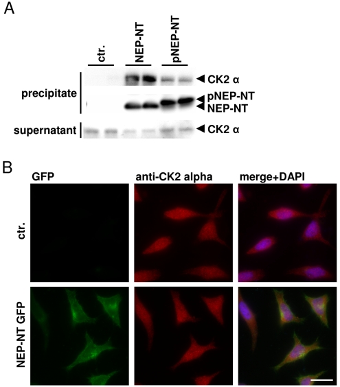
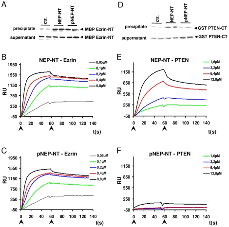
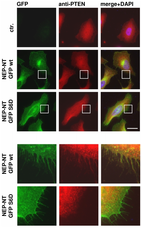
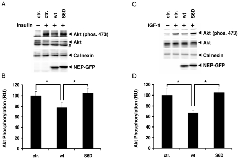
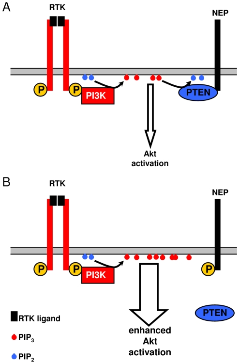
Neprilysin (NEP) is a type II membrane metalloproteinase that cleaves physiologically active peptides at the cell surface thus regulating the local concentration of these peptides available for receptor binding and signal transduction. In addition, the cytoplasmic N-terminal domain of NEP interacts with the phosphatase and tensin homologue deleted on chromosome 10 (PTEN) thereby regulating intracellular signaling via Akt. Thus, NEP serves dual functions in extracellular and intracellular signal transduction. Here, we show that NEP undergoes phosphorylation at serine residue 6 within the N-terminal cytoplasmic domain. In vitro and cell culture experiments demonstrate that Ser 6 is efficiently phosphorylated by protein kinase CK2. The phosphorylation of the cytoplasmic domain of NEP inhibits its interaction with PTEN. Interestingly, expression of a pseudophosphorylated NEP variant (Ser6Asp) abrogates the inhibitory effect of NEP on insulin/insulin-like growth factor-1 (IGF-1) stimulated activation of Akt. Thus, our data demonstrate a regulatory role of CK2 in the interaction of NEP with PTEN and insulin/IGF-1 signaling.


Conflict of interest statement

Competing Interests: The authors have declared that no competing interests exist.

Figures

Figure 1
Figure 1. NEP is phosphorylated by CK2 at serine 6.
A, Alignment of the N-terminal amino acid sequences of NEP from several mammalian species. Potential phosphorylation sites predicted by the NetPhos 2.0 server (with score >0.9) are indicated by asterisks. B, C, D HEK293 cells transiently expressing myc-tagged NEP wt or respective mutants were labeled with [32P] orthophosphate in presence or absence of okadaic acid (OA) (B) or DMAT (D) as indicated. After immunoprecipitation, the radiolabeled proteins were detected by autoradiography (upper panels). Subsequently, NEP was detected by immunoblotting with antibody 9E10 on the same membrane (lower panel). E, GST-fusion proteins representing amino acid 1–28 of the NEP N-terminus (GST-NEP) were incubated with CK2 in the presence of [γ-32P]ATP. The radiolabeled proteins were detected by autoradiography (upper panels). Subsequently, GST-fusion proteins were detected by immunoblotting with antibody against GST on the same membrane (lower panel). m = mature; im = immature; ctr. = untransfected cells.
Figure 2
Figure 2. Localization of full-length NEP and GFP-tagged chimeric proteins at the cell surface.
A, Schematic representation of full-length (fl) NEP and the chimeric NEP N-terminal GFP variant (NEP-NT GFP). B, Non-permeabilized HeLa cells expressing NEP-NT GFP wt or NEP-NT GFP Ser6Asp (S6D) were stained with mouse monoclonal antibody against GFP. The primary antibody was detected by Alexa 594-conjugated anti mouse secondary antibody. Images are representative for the typical distribution of NEP-GFP variants in several independent experiments. Note that NEP-NT GFP wt and the S6D Mutant are transported to the plasma membrane. C, HEK293 cells transiently expressing full-length NEP wt or NEP S6D and HEK293 cells stably expressing NEP-NT GFP wt or NEP-NT GFP S6D were cell surface biotin labeled. Biotinylated proteins were subsequently isolated with SA-agarose. All NEP variants are transported to the cell surface. Note the selective biotin labeling of the mature forms of full-length NEP. m = mature; im = immature; scale bar = 20 µm; ctr. = untransfected cells.
Figure 3
Figure 3. CK2 directly interacts with the N-terminus of NEP in vitro.
A, Biotin-labeled peptides that represent the N-terminal domain of non-phosphorylated (NEP-NT) or phosphorylated (pNEP-NT) NEP were coupled to SA-agarose beads and incubated with purified recombinant CK2. Precipitated proteins were detected by western immunoblotting. B, HeLa cells transiently expressing NEP-NT GFP were stained with mouse monoclonal antibody against CK2 alpha. The primary antibody was detected by Alexa 594-conjugated anti mouse secondary antibody. Scale bar = 20 µm; ctr. (A) = no peptide; ctr. (B) = untransfected cells.
Figure 4
Figure 4. Phosphorylation of NEP selectively inhibits the interaction with PTEN.
A, Biotin-labeled peptides that represent the N-terminal domain of non-phosphorylated (NEP-NT) or phosphorylated (pNEP-NT) NEP were coupled to SA-agarose beads and incubated with MBP Ezrin-NT (A) or GST PTEN-CT (D). SA-agarose precipitated proteins were detected by western immunoblotting. B, C, Sensograms of the interactions of MBP Ezrin-NT (concentrations ranging from 0.05 µM – 0.8 µM) with non-phosphorylated (NEP-NT; B) or phosphorylated (pNEP-NT; C) cytoplasmic domains of NEP. E, F, Sensograms of the interactions of GST PTEN-CT (concentrations ranging from 1.6 µM–12.8 µM) with non-phosphorylated (NEP-NT; E) or phosphorylated (pNEP-NT; F) cytoplasmic domains of NEP. Note that phosphorylation of NEP strongly inhibits the interaction with PTEN, while that with Ezrin is not affected. Arrows indicate start and end of injection, respectively; RU = response units; ctr. = no peptide.
Figure 5
Figure 5. Decreased interaction of pseudophsphorylated NEP with PTEN at the cell surface.
HeLa cells transiently expressing NEP-NT GFP wt or NEP-NT GFP S6D were stained with rabbit polyclonal antibody against PTEN and Alexa 594-conjugated anti rabbit secondary antibody. Note the selective co-localization of NEP-NT GFP wt and PTEN at the cell surface. Images are representative for the typical distribution of NEP-NT GFP variants and PTEN in several independent experiments. The boxed areas are enlarged in the lower panels. Scale bar = 20 µm; ctr. = untransfected cells.
Figure 6
Figure 6. Selective inhibition of Akt activation by non-phosphorylated NEP upon stimulation with insulin or IGF-1.
AD, HEK293 cells stably expressing NEP-NT GFP wt or NEP-NT GFP S6D cells were incubated for 16 hrs in serum free medium and then treated with 100 nM insulin (A, B) or 1 ng/ml IGF-1 (C, D) for 20 min. Non-transfected cells served as controls. Cells were lysed and proteins were detected by western immunoblotting. Quantification of the Akt phosphorylation (serine 473) upon stimulation with insulin (B) or IGF-1 (D) was done by ECL imaging. Calnexin signals were used as loading control. Values represent means ± S.D. (n = 3); * p-value<0.05. # = unspecific band; ctr. = untransfected cells.
Figure 7
Figure 7. Model for the role of NEP phosphorylation in Akt signalling.
PI3K is recruited to RTKs upon stimulation with specific ligands and phosphorylates PIP2 to PIP3. A, Non-phosphorylated NEP interacts with PTEN that antagonizes PI3K by the dephosphorylation of PIP3 to PIP2 resulting in the decreased Akt activation. B, The phosphorylation of NEP inhibits recruitment of PTEN to the plasma membrane and thereby stabilizes the pool of PIP3. Thus phosphorylation of NEP could increase or prolong the activation of Akt.

Similar articles

Cited by

References

    1. Kenny J. Endopeptidase-24.11: putative substrates and possible roles. Biochem Soc Trans. 1993;21(Pt 3):663–668. - PubMed
    1. Shipp MA, Tarr GE, Chen CY, Switzer SN, Hersh LB, et al. CD10/neutral endopeptidase 24.11 hydrolyzes bombesin-like peptides and regulates the growth of small cell carcinomas of the lung. Proc Natl Acad Sci U S A. 1991;88:10662–10666. - PMC - PubMed
    1. Iwata N, Tsubuki S, Takaki Y, Shirotani K, Lu B, et al. Metabolic regulation of brain Abeta by neprilysin. Science. 2001;292:1550–1552. - PubMed
    1. Hemming ML, Patterson M, Reske-Nielsen C, Lin L, Isacson O, et al. Reducing amyloid plaque burden via ex vivo gene delivery of an Abeta-degrading protease: a novel therapeutic approach to Alzheimer disease. PLoS Med. 2007;4:e262. - PMC - PubMed
    1. Stewart JR, Kenny AJ. Proteins of the kidney microvillar membrane. Biosynthesis of endopeptidase-24.11, dipeptidylpeptidase IV and aminopeptidases N and A in pig kidney slices. Biochem J. 1984;224:549–558. - PMC - PubMed

Publication types

MeSH terms