

Choroidal osteoma with choroidal neovascular membrane: Successful treatment with intravitreal bevacizumab
- PMID: 20957052
- PMCID: PMC2952608
- DOI: 10.2147/OPTH.S13730
Choroidal osteoma with choroidal neovascular membrane: Successful treatment with intravitreal bevacizumab
Abstract
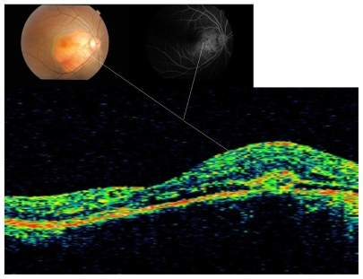
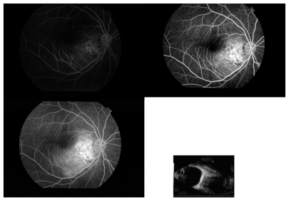
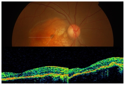
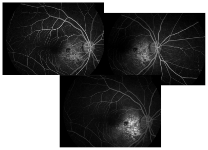
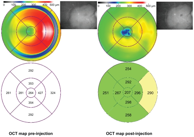
An otherwise healthy 27-year-old woman presented with complaints of sudden painless blurred vision in the right eye for one week. On examination, visual acuity was 20/30 in the right eye and 20/20 in left eye. Fundus examination OS was normal, but OD demonstrated an elevated, opaque, yellowish parapapillary choroidal lesion with grayish membrane associated with minimal subretinal fluid, suggestive of a choroidal neovascular membrane in the center. B-scan ultrasonography revealed findings consistent with a choroidal osteoma. Fundus fluorescein angiography of the right eye revealed a relatively well defined area of hyperfluorescence that increased in size and intensity in the later phases, suggestive of active extrafoveal choroidal neovascular membrane. Optical coherence tomography confirmed the extrafoveal choroidal neovascular membrane with subfoveal fluid. She was treated with intravitreal bevacizumab OD. At the two-week visit, vision OD improved to 20/20. Fluorescein angiography and optical coherence tomography revealed a resolved choroidal neovascular membrane. Intravitreal bevacizumab may be an effective alternative in the management of choroidal neovascular membrane secondary to choroidal osteoma.
Keywords: bevacizumab; choroidal neovascular membrane; optical coherence tomography; osteoma.
Figures
References
-
- Gass JD, Guerry RK, Jack RL, Harris G. Choroidal osteoma. Arch Ophthalmol. 1978;96:428–435. - PubMed
-
- Shields CL, Shields JA, Augsburger JJ. Choroidal osteoma. Surv Ophthalmol. 1988;33:17–27. - PubMed
-
- Aylward GW, Chang TS, Pautler SE, Gass JD. A long-term follow-up of choroidal osteoma. Arch Ophthalmol. 1998;116:1337–1341. - PubMed
-
- Kadrmas EF, Weiter JJ. Choroidal osteoma. Int Ophthalmol Clin. 1997;37:171–182. - PubMed
-
- Grand MG, Burgess DB, Singerman LJ, Ramsey J. Choroidal osteoma. Treatment of associated subretinal neovascular membranes. Retina. 1984;4:84–89. - PubMed
Publication types
LinkOut - more resources
Full Text Sources
