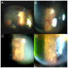
Transient tractional retinal detachment in an eye with retinitis pigmentosa
- PMID: 20957057
- PMCID: PMC2952613
- DOI: 10.2147/OPTH.S13885
Transient tractional retinal detachment in an eye with retinitis pigmentosa
Abstract
We present a case of retinitis pigmentosa with vitreoretinal traction-associated retinal detachment. The retinal detachment was detected in the nasal periphery. No retinal breaks and no active vascular leakage were observed by fundus scopy and fluorescein angiography, respectively. However, 8 months later, the tractional retinal detachment was spontaneously resolved with posterior vitreous detachment.
Keywords: retinal detachment; retinitis pigmentosa; vitreoretinal traction.
Figures


References
-
- Fishman GA, Fishman M, Maggiano J. Macular lesions associated with retinitis pigmentosa. Arch Ophthalmol. 1977;95:798–803. - PubMed
-
- Wise GN. Clinical features of idiopathic preretinal macular fibrosis. Schoenberg Lecture. Am J Ophthalmol. 1975;79:349–357. - PubMed
-
- Demir MN, Unl N, Yalniz Z, Acar MA, Ornek F. A case of retinal detachment in retinitis pigmentosa. Eur J Ophthalmol. 2007;17:677–679. - PubMed
-
- Rani A, Pal N, Azad RV, Sharma YR, Chandra P, Vikram Singh D. Tractional retinal detachment in usher syndrome type II. Clin Experiment Ophthalmol. 2005;33:436–437. - PubMed
-
- Hong PH, Han DP, Burke JM, Wirostko WJ. Vitrectomy for large vitreous opacity in retinitis pigmentosa. Am J Ophthalmol. 2001;131:133–134. - PubMed
Publication types
LinkOut - more resources
Full Text Sources

