Review
. 2010 Oct 4;2(5):e00045.
doi: 10.1042/AN20100019.

Mitochondria and neuroplasticity

Affiliations
Review

Mitochondria and neuroplasticity

Aiwu Cheng et al. ASN Neuro. .

Abstract

The production of neurons from neural progenitor cells, the growth of axons and dendrites and the formation and reorganization of synapses are examples of neuroplasticity. These processes are regulated by cell-autonomous and intercellular (paracrine and endocrine) programs that mediate responses of neural cells to environmental input. Mitochondria are highly mobile and move within and between subcellular compartments involved in neuroplasticity (synaptic terminals, dendrites, cell body and the axon). By generating energy (ATP and NAD(+)), and regulating subcellular Ca(2+) and redox homoeostasis, mitochondria may play important roles in controlling fundamental processes in neuroplasticity, including neural differentiation, neurite outgrowth, neurotransmitter release and dendritic remodelling. Particularly intriguing is emerging data suggesting that mitochondria emit molecular signals (e.g. reactive oxygen species, proteins and lipid mediators) that can act locally or travel to distant targets including the nucleus. Disturbances in mitochondrial functions and signalling may play roles in impaired neuroplasticity and neuronal degeneration in Alzheimer's disease, Parkinson's disease, psychiatric disorders and stroke.

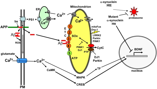
Keywords: AD, Alzheimer's disease; AP, adaptor protein; APP, amyloid precursor protein; Aβ, amyloid β-peptide; BDNF, brain-derived neurotrophic factor; CR, caloric restriction; CREB, cAMP-response-element-binding protein; CaMK, Ca2+/calmodulin-dependent protein kinase; ES, embryonic stem; ETC, electron transport chain; HD, Huntington's disease; LRRK2, leucine-rich repeat kinase 2; LTP, long-term potentiation; MAPK, mitogen-activated protein kinase; Mn-SOD, manganese superoxide dismutase; NGF, nerve growth factor; NMDA, N-methyl-d-aspartate; Nrf1, nuclear respiratory factor 1; OPA1, Optic Atrophy-1; PD, Parkinson's disease; PGC1α, peroxisome-proliferator-activated receptor γ co-activator 1α; PINK1, PTEN (phosphatase and tensin homologue deleted on chromosome 10)-induced kinase 1; PPAR, peroxisome-proliferator-activated receptor; UCP, uncoupling protein; mitochondria biogenesis; mitochondria fission and fusion; neural progenitor cell.


Figures

Figure 1
Figure 1. Examples of methods for evaluating mitochondrial morphology, subcellular localization and functional status
(a) Each panel shows a single live embryonic rat hippocampal neuron at a different stage of development in culture (1, 5 and 14 days). Mitochondria in the neurons were stained with the fluorescent probe MitoTracker Red. Note that the immature neuron has elaborated short processes, and that the vast majority of mitochondria are clustered in a perinuclear location. At 5 days in culture the neuron exhibits longer neurites that contain multiple mitochondria, and perinuclear mitochondria remain abundant. Mitochondrial numbers have increased, indicating that biogenesis has occurred. At 14 days in culture the neuron has elaborated an extensive neuritic network with each neurite containing multiple mitochondria. Relative numbers of mitochondria in a perinuclear location are reduced. (b) Example of the results of an experiment in which oxygen consumption, mitochondrial membrane potential and cytoplasmic Ca2+ levels were monitored in a single cultured embryonic rat hippocampal neuron before and during exposure to the excitatory neurotransmitter glutamate. In response to glutamate receptor activation, oxygen consumption increased and then slowly returned towards baseline levels, intracellular Ca2+ levels rose rapidly and remained elevated, whereas mitochondrial membrane potential declined progressively. Adapted from Gleichmann et al. (2009).
Figure 2
Figure 2. Molecular machinery that actively moves mitochondria to and fro within axons
A major mechanism by which mitochondria are transported in either anterograde or retrograde directions in axons involves their energy (ATP)-dependent movement along microtubules. ATP-dependent ‘motor’ proteins interact with the microtubules to generate the force that moves the mitochondria in anterograde (kinesin) or retrograde (dynein) directions respectively. Several APs (adaptor proteins) mediate the interaction of mitochondria with motor proteins, including APs that interact with kinesin (Milton, syntabulin and the Rho GTPase Miro) and APs that associate with dynein (dynactin). In addition, in synaptic terminals and growth cones, microtubules may be moved by myosin-mediated interactions with actin filaments. Myosin V can drive short-range movements along F-actin, as well as modulate long-range transport by pulling mitochondria away from microtubules by facilitating anchorage of mitochondria to F-actin by unknown actin–mitochondrion crosslinkers. Adapted from Mattson et al. (2008).
Figure 3
Figure 3. The landscape of mitochondrial involvement in the plasticity of neuronal structure and information processing
Increasing evidence suggests that mitochondria play active roles in regulating the outgrowth of axons and dendrites, synaptogenesis and morphological and functional responses to synaptic activity. Mitochondria in presynaptic terminals (1) provide the energy for the maintenance and restoration of membrane potential, and may modulate neurotransmitter packaging and release. Mitochondria in postsynaptic spines (2) and dendritic shafts (3) may enable/regulate both structural and functional responses of these compartments to synaptic activity. Mitochondria in the cell body (4) provide the energy required for numerous biochemical processes, and may also serve as signalling platforms involved in information transfer within the neuron. Mitochondria in axons (5) provide the energy necessary for the transport of various proteins and organelles from the axon terminal to the cell body and vice versa.
Figure 4
Figure 4. Mechanisms that regulate the growth and function of mitochondria
The vast majority of mitochondrial proteins are encoded by nuclear genes, whereas only 13 proteins that are all components of the ETC are encoded by mitochondrial genes. Proteins are imported into mitochondria by translocases of the outer membrane (TOM) and inner membrane (TIM) in a mitochondrial membrane potential-dependent manner. Mitochondrial DNA transcription is mediated by an RNA polymerase, the transcription factor A (TFAM), TFB1M or TFB2M for transcription initiation, and mTERE for transcription termination. Neurons contain particularly high levels of PGC1α, apparently because of their high energy utilization. PGC1α is a transcriptional co-activator that interacts with various transcription factors, including the nuclear receptors PPARγ, PPARα, oestrogen, thyroid hormone and retinoid receptors and Nrf1/2. Nrf1/2 induces the transcription of nuclear genes that encode ETC proteins, TFAM and proteins critical for mitochondrial biogenesis. Environmental factors (exercise, energy restriction and cold temperature, for example) activate signalling pathways involving neurotransmitters, neurotrophic factors and nitric oxide (for example), and intracellular cascades involving Ca2+, CaMKII (Ca2+/calmodulin-dependent protein kinase II), the phosphatase calcineurin, and the transcription factor CREB (cAMP-response-element-binding protein). In this manner, PGC1α is activated and mitochondrial biogenesis is stimulated. Modified from Mattson et al. (2008).
Figure 5
Figure 5. Influence of neurodegenerative disease-related proteins proteins on mitochondrial function and plasticity
The AD APP is a single-pass transmembrane protein that includes the 42-amino-acid Aβ (red). Aβ is liberated upon sequential cleavages of APP by β-secretase (bs) at the N-terminus of Aβ and γ-secretase at the C-terminus of Aβ; the latter cleavage is executed by presenilin-1 (PS1; an integral membrane protein that is the enzymatic component of the γ-secretase protein complex). An alternative cleavage of APP within the Aβ sequence by the α-secretase enzyme produces a secreted form of APP that is believed to play important roles in developmental and synaptic plasticity, and neuron survival. Whereas Aβ is normally cleared from the brain, in AD it self-aggregates to form oligomers and during this process ROS are generated and propagated to the cell membrane, resulting in lipid peroxidation, impairment of membrane ion (Na+ and Ca2+) transporters and excessive Ca2+ influx through glutamate receptor (NMDA) channels. In addition to increasing Aβ production, mutations in PS1 that cause AD perturb endoplasmic reticulum (ER) Ca2+, resulting in excessive Ca2+ release. Mitochondrial Ca2+ homoeostasis and energy production may be impaired in neurons in AD as the result of cellular Ca2+ overload and increased oxidative stress possibly involving direct actions of Aβ on mitochondrial membranes. The pathological scenario just described involves perturbations in signalling pathways normally involved in adaptive neuroplasticity, with glutamate signalling being a well-established example. Glutamate-induced Ca2+ is normally transient with the Ca2+ activating kinases such as CaMK and MAPK. The kinases in turn activate transcription factors (TF) that induce the expression of nuclear genes that encode proteins involved in effecting adaptive morphological and functional responses of the neurons. CREB is one such synaptic activity-responsive transcription factor that induces the expression of BDNF, a protein critically involved in synaptic plasticity and neuronal survival. The successive discoveries of genes-linked inherited forms of PD placed mitochondrial alterations at centre stage in the pathogenesis of PD, and also revealed novel proteins that regulate mitochondrial functional and structural plasticity. A feature of all cases of PD is the intracellular accumulation of α-synuclein, apparently as the result of impaired degradation by the proteasome. Parkin (an E3 ubiquitin ligase), DJ-1, PINK1 and LRRK2 have each been shown to modify mitochondrial function (ATP and ROS production), and some findings suggest that Parkin, PINK1 and LRRK2 also influence mitochondrial fission and fusion. PINK1 and DJ-1 may suppress intramitochondrial oxidative stress, whereas Parkin and DJ-1 inhibit the opening of mitochondrial permeability transition pores, thereby protecting neurons against apoptotic cell death that can be triggered by release of cytochrome c (CytC) from the mitochondria. Finally, in HD, mutant huntingtin (Htt) self-aggregates and impairs CREB-mediated transcription of BDNF, thereby compromising a pathway critical for neuronal plasticity and survival. See the text of this paper and the following references for additional information and discussion (Mattson, 1997; Mattson, 2004; Mattson et al., 2004; Cattaneo et al., 2005; Cookson, 2005; Nuytemans et al., 2010).

Similar articles

Cited by

References

    1. Adhihetty PJ, Beal MF. Creatine and its potential therapeutic value for targeting cellular energy impairment in neurodegenerative diseases. Neuromolecular Med. 2008;10:275–290. - PMC - PubMed
    1. Andersson U, Scarpulla RC. Pgc-1-related coactivator, a novel, serum-inducible coactivator of nuclear respiratory factor 1-dependent transcription in mammalian cells. Mol Cell Biol. 2001;21:3738–3749. - PMC - PubMed
    1. Auffret A, Gautheron V, Mattson MP, Mariani J, Rovira C. Progressive age-related impairment of the late long-term potentiation in Alzheimer's disease presenilin-1 mutant knock-in mice. J Alzheimers Dis. 2010;19:1021–1033. - PMC - PubMed
    1. Baar K, Wende AR, Jones TE, Marison M, Nolte LA, Chen M, Kelly DP, Holloszy JO. Adaptations of skeletal muscle to exercise: rapid increase in the transcriptional coactivator PGC-1. FASEB J. 2002;16:1879–1886. - PubMed
    1. Barres BA. The mystery and magic of glia: a perspective on their roles in health and disease. Neuron. 2008;60:430–440. - PubMed