. 2010 Sep 2;11(9):3106-21.
doi: 10.3390/ijms11093106.

Identification of tetranectin as a potential biomarker for metastatic oral cancer

Affiliations

Identification of tetranectin as a potential biomarker for metastatic oral cancer

Martha E Arellano-Garcia et al. Int J Mol Sci. .

Abstract

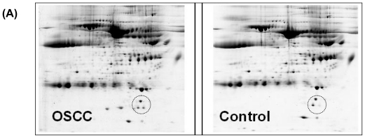
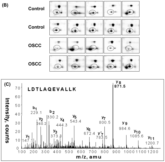
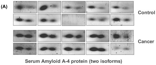
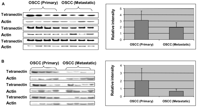
Lymph node involvement is the most important predictor of survival rates in patients with oral squamous cell carcinoma (OSCC). A biomarker that can indicate lymph node metastasis would be valuable to classify patients with OSCC for optimal treatment. In this study, we have performed a serum proteomic analysis of OSCC using 2-D gel electrophoresis and liquid chromatography/tandem mass spectrometry. One of the down-regulated proteins in OSCC was identified as tetranectin, which is a protein encoded by the CLEC3B gene (C-type lectin domain family 3, member B). We further tested the protein level in serum and saliva from patients with lymph-node metastatic and primary OSCC. Tetranectin was found significantly under-expressed in both serum and saliva of metastatic OSCC compared to primary OSCC. Our results suggest that serum or saliva tetranectin may serve as a potential biomarker for metastatic OSCC. Other candidate serum biomarkers for OSCC included superoxide dismutase, ficolin 2, CD-5 antigen-like protein, RalA binding protein 1, plasma retinol-binding protein and transthyretin. Their clinical utility for OSCC detection remains to be further tested in cancer patients.

Keywords: disease biomarker; oral squamous cell carcinoma; serum proteomics; tetranectin.


Figures

Figure 1
Figure 1
Analysis of serum proteins from OSCC and healthy control subjects using 2-DE with tandem MS. (A) 2-DE separation of serum proteins. 1st-dimension separation: IEF, IPG strips (pI 3–10, non-linear. 2nd-dimension: SDS-PAGE, 18-cm 8–16% gradient gels. The proteins were visualized using Sypro Ruby staining; (B) Serum tetranectin (circled) was found at significantly reduced level in OSCCs (n = 10) compared to healthy controls (n = 10), based on 2-DE analysis; (C) ESI-MS/MS spectrum of a tryptic peptide, LDTLAQEVALLK, originated from tetranectin. The protein was identified by using in-gel digestion and LC-MS/MS analysis of the resulting peptides.
Figure 1
Figure 1
Analysis of serum proteins from OSCC and healthy control subjects using 2-DE with tandem MS. (A) 2-DE separation of serum proteins. 1st-dimension separation: IEF, IPG strips (pI 3–10, non-linear. 2nd-dimension: SDS-PAGE, 18-cm 8–16% gradient gels. The proteins were visualized using Sypro Ruby staining; (B) Serum tetranectin (circled) was found at significantly reduced level in OSCCs (n = 10) compared to healthy controls (n = 10), based on 2-DE analysis; (C) ESI-MS/MS spectrum of a tryptic peptide, LDTLAQEVALLK, originated from tetranectin. The protein was identified by using in-gel digestion and LC-MS/MS analysis of the resulting peptides.
Figure 2
Figure 2
Serum amyloid A-4 protein (SAA-4) is significantly over-expressed in OSCC compared to healthy individuals. (A) Both isoforms of SAA-4 were found significantly higher in OSCC than healthy controls, based on 2-DE analysis; (B) The ESI-MS/MS spectrum for a tryptic peptide, EALQGVGDMGR, derived from SAA-4.
Figure 2
Figure 2
Serum amyloid A-4 protein (SAA-4) is significantly over-expressed in OSCC compared to healthy individuals. (A) Both isoforms of SAA-4 were found significantly higher in OSCC than healthy controls, based on 2-DE analysis; (B) The ESI-MS/MS spectrum for a tryptic peptide, EALQGVGDMGR, derived from SAA-4.
Figure 3
Figure 3
(A) Western blot analysis of tetranectin in serum samples from primary (n = 12) and lymph-node metastatic (n = 12) OSCC subjects; (B) Western blotting of tetranectin in saliva samples from primary OSCC (n = 12) and lymph-node metastatic OSCC (n = 12). The bar figures show the normalized level (y-axis) of tetranectin against actin. Tetranectin was significantly under-expressed in metastatic versus primary cancer (Serum: p = 0.03; saliva: p = 0.007).

Similar articles

Cited by

References

    1. Argiris A, Karamouzis MV, Raben D, Ferris RL. Head and neck cancer. Lancet. 2008;371:1695–1709. - PMC - PubMed
    1. Parkin DM, Bray F, Ferlay J, Pisani P. Global Cancer Statistics, 2002. CA: Cancer J. Clin. 2005;55:74–108. - PubMed
    1. Grandi C, Alloisio M, Moglia D, Podrecca S, Sala L, Salvatori P, Molinari R. Prognostic significance of lymphatic spread in head and neck carcinomas: Therapeutic implications. Head Neck Surg. 1985;8:67–73. - PubMed
    1. Greenberg JS, Fowler R, Gomez J, Mo V, Roberts D, Naggar AKE, Myers JN. Extent of extracapsular spread. Cancer. 2003;97:1464–1470. - PubMed
    1. Sano D, Myers J. Metastasis of squamous cell carcinoma of the oral tongue. Cancer Metastasis Rev. 2007;26:645–662. - PubMed

Publication types