. 2010 Oct 5;5(10):e13183.
doi: 10.1371/journal.pone.0013183.

VCP associated inclusion body myopathy and paget disease of bone knock-in mouse model exhibits tissue pathology typical of human disease

Affiliations

VCP associated inclusion body myopathy and paget disease of bone knock-in mouse model exhibits tissue pathology typical of human disease

Mallikarjun Badadani et al. PLoS One. .

Abstract

Dominant mutations in the valosin containing protein (VCP) gene cause inclusion body myopathy associated with Paget's disease of bone and frontotemporal dementia (IBMPFD). We have generated a knock-in mouse model with the common R155H mutation. Mice demonstrate progressive muscle weakness starting approximately at the age of 6 months. Histology of mutant muscle showed progressive vacuolization of myofibrils and centrally located nuclei, and immunostaining shows progressive cytoplasmic accumulation of TDP-43 and ubiquitin-positive inclusion bodies in quadriceps myofibrils and brain. Increased LC3-II staining of muscle sections representing increased number of autophagosomes suggested impaired autophagy. Increased apoptosis was demonstrated by elevated caspase-3 activity and increased TUNEL-positive nuclei. X-ray microtomography (uCT) images show radiolucency of distal femurs and proximal tibiae in knock-in mice and uCT morphometrics shows decreased trabecular pattern and increased cortical wall thickness. Bone histology and bone marrow derived macrophage cultures in these mice revealed increased osteoclastogenesis observed by TRAP staining suggestive of Paget bone disease. The VCP(R155H/+) knock-in mice replicate the muscle, bone and brain pathology of inclusion body myopathy, thus representing a useful model for preclinical studies.


Conflict of interest statement

Competing Interests: The authors have declared that no competing interests exist.

Figures

Figure 1
Figure 1. Generation of the VCPR155H/+ knock-in mice.
(A) Schematic drawing of the R155H targeting strategy of the knock-in (KI) allele. The top line shows the knock-in (KI) allele and the below line depicts the wild-type (WT) allele. The localizations of exons 1 through 5 are numbered. Localizations of the 5′ (7.9 kb) and 3′ (2.1 kb) targeting sequences are indicated by dashed lines. Neomycin-cassette is marked by Neo, flanked by FRT sites and the restriction enzyme sites are indicated as follows: K  =  Kpn I, Bg  =  Bgl II, R  =  EcoR I, H  =  Hind III. Black arrow heads flanking Neo cassette show LoxP sites and white arrow heads show FRT sites. Black arrows flanking exon 5 indicate the locations of primers used in genotyping. (B) PCR genotyping of isolated from mouse tail DNA and digested by Msp I produces 274 bp and 362 bp in wild-type, while mutant allele gives three fragments of 636 bp, 274 bp and 362 bp. (C) RT-PCR-Msp I digestion analysis of mRNA isolated from mouse quadriceps shows similar expression levels. Wild-type allele produces 700 bp and 282 bp fragments, and mutant allele produces a 982 bp fragment. Fragment sizes are shown on the right and genotypes above. (D) VCP expression by western immunoblot in tissues: quadriceps, brain, heart and liver in wild type compared with knock-in mouse model shows similar expression levels. (E) VCP expression by Western immunoblot in quadriceps from wild type compared with knock-in mouse model. Beta actin was used as a loading control for these western blot analyses.
Figure 2
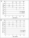
Figure 2. Progression of muscle weakness of the quadricep muscles in knock-in mice.
(A) Decline of physical performance by Rotarod analysis in knock-in mice. (B) Progressive impairment of muscle strength measured by grip strength meter in knock-in mice. Ages of mice are shown in the X-axis and results are indicated in the Y-axis as relative values when compared to the wild-type mouse values. The following numbers of mice have been used to analyze physical performance test at different time points: 3 months (25 wild-type, 9 knock-in), 6 months (51 wild-type, 20 knock-in), 9 months (48 wild-type, 18 knock-in), 12 months (24 wild-type, 9 knock-in), and 15 months (20 wild-type, 8 knock-in). Note * * in the figure represents p value <0.05.
Figure 3
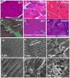
Figure 3. Immunohistochemical analysis in the wild-type and VCPR155H/+ knock-in mouse muscle.
(A–C) Quadricep muscles from 9–10 month-old wild-type and VCPR155H/+ knock-in mice were analyzed by H&E staining. (B) An enlarged vacuole in the mutant tissue is shown by white arrows and (C) centrally located nuclei and rimmed vacuoles are revealed in the mutant mice shown by white arrows. (D) Quadriceps muscles from 15-month old VCPR155H/+ knock-in mice, centrally located nuclei shown by white arrows. (E–F) Modified Gomori Trichrome staining of muscle fibers from wild type and VCPR155H/+ knock-in mice. Magnification: 630×. (G–L) Electron microscopy analyses of the mouse quadricep muscles. Vacuolization and loss of myofilament organization are observed in quadriceps muscle from 10-month-old VCPR155H/+ knock-in mice (G–I), but not in wild-type mice (J–L). Sarcomeric direction is indicated by white double ended arrow (H,K). Swollen mitochondria are also observed in the mutant tissue (L). Black arrows in (K) and (L) indicate accumulation of vacuoles. Size bars are shown in the lower left corner of each image. Mt  =  mitochondria. Magnifications: E+H  = 900×, F+I  = 2,950×, G+J  = 11,500×. (N = 3 WT and 3 R155H animals).
Figure 4
Figure 4. Immunohistochemical analysis and protein expression in the wild-type and VCPR155H/+ knock-in mouse muscle.
(A–F) Immunohistochemical analysis of quadriceps muscles from 9–10 month old wild-type (A–C) and VCPR155H/+ knock-in mice (D–F) were stained with a ubiquitin-specific FK1 antibody (A,D) and a TDP-43-specific antibody (B,E). (C) shows the overlay of (A) and (B), and (F) is the overlay of (D) and (E). Ubiquitin- and TDP-43-positive, cytoplasmic inclusion body is shown by an arrow in (D–F). Nuclei were stained with DAPI. Magnification: 630×.). (G) Expression of TDP-43 and ubiquitinated proteins. Proteins were harvested from the quadriceps muscle of 2 littermates of wild-type and knock-in mice and analyzed by Western blotting using TDP-43 (upper panel) and ubiquitin/FK1 (lower panel) antibodies. Each membrane was re-probed with actin to confirm equal protein loading in each lane. Protein bands are indicated on the left and molecular weights of marker bands for the ubiquitin blot on the right. Genotypes are shown above. Wild-type and knock-in samples are from two litters (indicated above the figure) (N = 4 WT and 4 R155H animals).
Figure 5
Figure 5. LC3-II staining, protein expression and apoptosis detection in the wild-type and VCPR155H/+ knock-in mouse quadriceps.
(A–B) Quadricep muscles from 9–10 month old wild type and VCPR155H/+ knock-in mice were stained with an LC3-II-specific antibody. Nuclei were stained with DAPI (Magnification: 630×). (C) Protein expression of LC3-II is increased in knock in mice as compared with wild type litter mates. (D–E) DAPI and TUNEL staining of the quadriceps section from a wild-type and VCPR155H/+ knock-in mouse models (N = 3 WT and 3 R155H animals). Apoptotic nuclei of the mutant tissue are shown by white arrows. Magnification: 400×. (F) Caspase-3 activity was measured from the quadriceps muscle lysates of wild-type and VCPR155H/+ knock-in mice. Specific activities are shown in the Y-axis and genotypes in the X-axis (N = 4 WT and 4 R155H animals).
Figure 6
Figure 6. Bone micro CT imaging, histology and OCL formation of VCPR155H/+ and wild-type mice.
(A–B) micro CT images showing sclerotic lesions at anterior tibia and posterior femur shown by white arrows in 15-month old knock-in mice. (C–D) Transverse sections of decalcified 6th lumbar vertebra, white arrowheads indicate red colored TRAP-positive osteoclasts (Magnification 10×). (E–G) OCLs formed from non-adherent marrow cell cultures for WT or VCPR155H/+ cultured for 9 days in the presence of 10 ng/ml M-CSF and varying concentrations of RANKL. The cells were then fixed and stained for TRAP activity. Results represent TRAP positive cells containing ≥ three nuclei and are expressed as the mean ± SEM for duplicate cultures.*P<0.05 compared with results from wt cell cultures. MNC =  multi-nuclear cell. (G) Large TRAP positive multinuclear OC-like cell from knock-in bone marrow derived macrophages (BMDM). Differentiated in 100 ng/ml RANKL and 50 ng/ml M-CSF then fixed and TRAP stained on day 9 of differentiation. (H) Osteoclastogenesis cytokine sensitivity was determined with increasing concentrations of RANKL. TRAP-expressing multi-nucleated OCs was scored (N = 4 WT and 4 R155H animals).
Figure 7
Figure 7. Immunohistochemical brain staining of the wild-type and R155H knock-in mouse.
(A–B) H&E staining of 15 month old wild type and knock-in mouse of hippocampus and neuronal injury shown by white arrows (Magnification 400×). (C–D) IBA1 staining of frontal cortex from wild-type and R155H knock-in mice shown by white arrows Magnification: 630× (N = 3 WT and 3 R155H animals).
Figure 8
Figure 8. Immunohistochemical staining and protein expression of the wild-type and R155H knock-in mouse brain.
(A–B) Co-localization of VCP- and ubiquitin-specific antibodies in 15-month old wild type and R155H knock-in mice. (C–D) Co-localization of FK1- and TDP-43-specific antibodies in wild type and R155H knock-in mice from frontal cortex. Inclusion bodies are shown by white arrows. Nuclei were stained with DAPI (blue). Magnification: 630× (N = 3 WT and 3 R155H animals). (E–F) Co-localization of VCP- and TDP-43-specific antibodies of wild type and R155H knock-in mice from frontal cortex. Inclusion bodies are positive for VCP and TDP-43 antibodies are shown by white arrows and inset. Nuclei were stained with DAPI. (G–H) Protein expression of VCP, TDP-43, GFAP, PUMA, BAX and ubiquitin have been analyzed by Western immunoblot from WT and R155H KI mutant whole brain extracts (N = 4 WT and 4 R155H animals). Beta actin was used as loading controls for these Western blot analyses.

References

    1. Kimonis VE, Kovach MJ, Waggoner B, Leal S, Salam A, et al. Clinical and molecular studies in a unique family with autosomal dominant limb-girdle muscular dystrophy and Paget disease of bone. Genet Med. 2000;2:232–241. - PMC - PubMed
    1. Kimonis VE, Mehta SG, Fulchiero EC, Thomasova D, Pasquali M, et al. Clinical studies in familial VCP myopathy associated with Paget disease of bone and frontotemporal dementia. Am J Med Genet A. 2008;146:745–757. - PMC - PubMed
    1. Kovach MJ, Waggoner B, Leal SM, Gelber D, Khardori R, et al. Clinical delineation and localization to chromosome 9p13.3-p12 of a unique dominant disorder in four families: hereditary inclusion body myopathy, Paget disease of bone, and frontotemporal dementia. Mol Genet Metab. 2001;74:458–475. - PMC - PubMed
    1. Watts GD, Thorne M, Kovach MJ, Pestronk A, Kimonis VE. Clinical and genetic heterogeneity in chromosome 9p associated hereditary inclusion body myopathy: exclusion of GNE and three other candidate genes. Neuromuscul Disord. 2003;13:559–567. - PubMed
    1. Kimonis V, Watts G. Inclusion Body Myopathy Associated with Paget Disease of Bone and/or Frontotemporal Dementia Gene GeneTests (wwwgenetestsorg) and University of Washington, Seattle 2007.

Publication types