. 2010 Oct 1;5(10):e13127.
doi: 10.1371/journal.pone.0013127.

Viewing ageing eyes: diverse sites of amyloid Beta accumulation in the ageing mouse retina and the up-regulation of macrophages

Affiliations

Viewing ageing eyes: diverse sites of amyloid Beta accumulation in the ageing mouse retina and the up-regulation of macrophages

Jaimie Hoh Kam et al. PLoS One. .

Abstract

Background: Amyloid beta (Aβ) accumulates in the ageing central nervous system and is associated with a number of age-related diseases, including age-related macular degeneration (AMD) in the eye. AMD is characterised by accumulation of extracellular deposits called drusen in which Aβ is a key constituent. Aβ activates the complement cascade and its deposition is associated with activated macrophages. So far, little is known about the quantitative measurements of Aβ accumulation and definitions of its relative sites of ocular deposition in the normal ageing mouse.

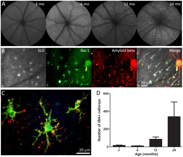
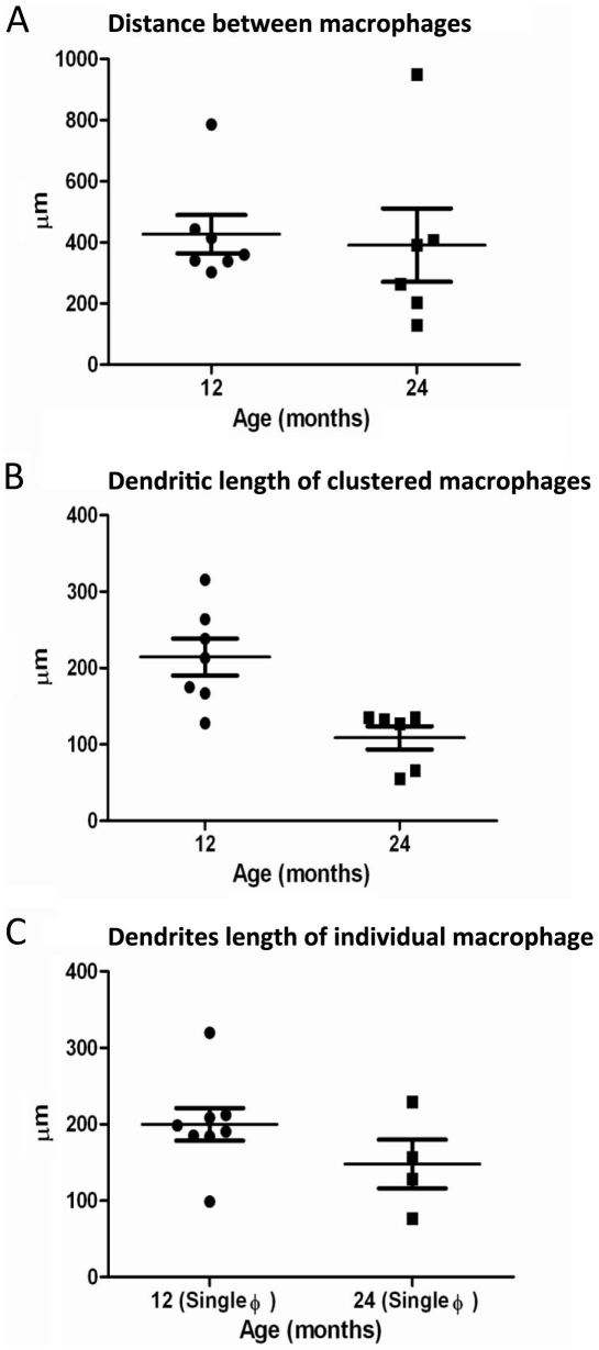
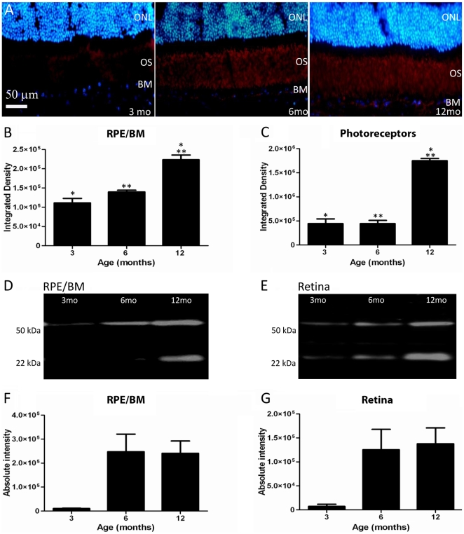
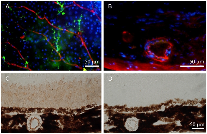
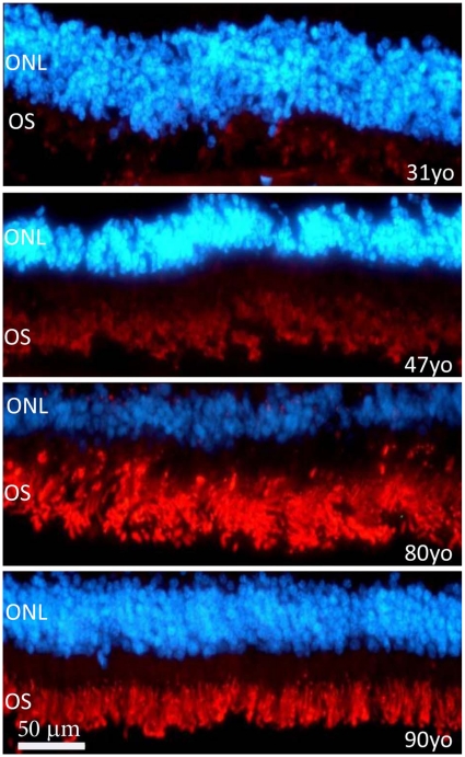
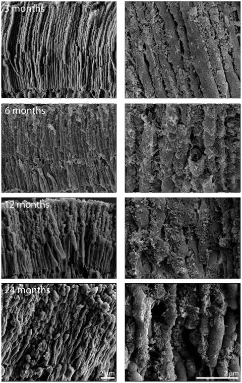
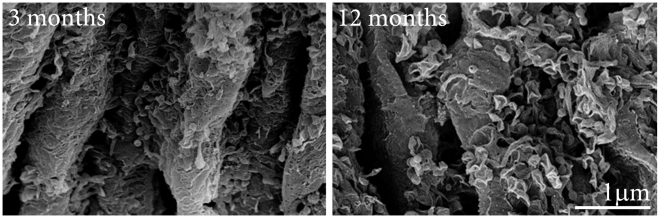
Methodology/principal findings: We have traced Aβ accumulation quantitatively in the ageing mouse retina using immunohistochemistry and Western blot analysis. We reveal that it is not only deposited at Bruch's membrane and along blood vessels, but unexpectedly, it also coats photoreceptor outer segments. While Aβ is present at all sites of deposition from 3 months of age, it increases markedly from 6 months onward. Progressive accumulation of deposits on outer segments was confirmed with scanning electron microscopy, revealing age-related changes in their morphology. Such progress of accumulation of Aβ on photoreceptor outer segments with age was also confirmed in human retinae using immunohistochemistry. We also chart the macrophage response to increases in Aβ showing up-regulation in their numbers using both confocal laser imaging of the eye in vivo followed by in vitro immunostaining. With age macrophages become bloated with cellular debris including Aβ, however, their increasing numbers fail to stop Aβ accumulation.

Conclusions: Increasing Aβ deposition in blood vessels and Bruch's membrane will impact upon retinal perfusion and clearance of cellular waste products from the outer retina, a region of very high metabolic activity. This accumulation of Aβ may contribute to the 30% reduction of photoreceptors found throughout life and the shortening of those that remain. The coating of Aβ on outer segments may also have an impact upon visual function with age.


Conflict of interest statement

Competing Interests: The authors have declared that no competing interests exist.

Figures

Figure 1
Figure 1. Retinal imaging and macrophage histology.
A. Scanning laser ophthalmoscope images of the retinae of mice taken at 3, 6, 12 and 24 months of age. With time there is an accumulation of fluorescent point sources. B. The images are overlaid with the eyecups of a 12 months old mouse once they have been stained for Iba-1 to reveal macrophages and Aβ. This shows that many of the point sources are macrophages containing Aβ (indicated with arrows). These are arranged in a grid like pattern. C. A higher power image of Aβ containing macrophages of a 12 months old mouse. D. The number of macrophages present in the whole mounts at progressive ages. There are significant increases at 12 and 24 months (see text for statistics).
Figure 2
Figure 2. Graphs showing the distance between macrophages and measurement of the dendritic processes.
A. Graph showing the distance between macrophages in a cluster of seven cells for the 12 months and six cells for the 24 months old mouse. B. Graph showing the length of the dendritic processes of these seven cells of the 12 months and six cells of the 24 months old mouse (P<0.01). C. Graph showing the length of the dendritic processes of eight individual macrophage cells for the 12 months and four individual cells for the 24 months old mouse.
Figure 3
Figure 3. Aβ is deposited at the Bruch's membrane (BM)/RPE interface and among photoreceptor outer segments.
A. The accumulation of amyloid beta in sections showing BM/RPE interface and the regions of the outer segments (OS) in mice of 3, 6 and 12 months age. Here Aβ label is red and the outer nuclear layer (ONL) is blue. This progressive accumulation was quantified with two independent methods at the two sites. First, the integrated density of label from immunostained sections was measured. The results of this are shown graphically in B and C. There are significant increases at both sites, particularly at 12 months (see text for levels of significance). Second, Western blots were run for Aβ at each site shown in D and E. F and G show the measurements at the same three time points as in B and C. The amount of Aβ increases significantly over time (see text for levels of significance). Differences between B and C and F and G are probably due to the different amounts of tissue sampled as F and G will also include measures derived from inner and outer retinal blood vessels.
Figure 4
Figure 4. Retinal blood vessels stained for Aβ.
A. Inner retinal vessels stained for Aβ (red), Iba-1 (green) and neuronal cell bodies (blue) at 24 months. The amyloid deposits can be seen to be at focal points along the vessel rather than being continuous. B. Choroidal vessels also accumulated Aβ, however, the accumulation of this material appeared to be specific to a sub-group of vessels with other showing no sign of Aβ accumulation. This is taken from a 12 months old animal. C. Immunostaining of retinal section of a 12 months old animal using the colorimetric method (3,3-diaminobenzidine) to confirm the presence of Aβ in the outer segment of the photoreceptor and in the blood vessels. D. Negative control of the colorimetric staining showing the absence of staining.
Figure 5
Figure 5. Aβ staining in human outer retina from individuals aged 31, 47, 80 and 90 years.
This was undertaken on retinae separated from the RPE. Aβ is red and the outer nuclear layer (ONL) is blue. The outer segments (OS) are positive for Aβ but the intensity of the staining increases with age. In spite of this, the overall progression of Aβ accumulation here mirrors that found in mice.
Figure 6
Figure 6. Scanning electron micrographs of photoreceptor outer segments taken from animals at 3, 6, 12 and 24 months of age.
In each case the right hand panel is a higher magnification of that on the left and the orientation is such that the RPE would be to the top and the outer nuclear layer to the bottom. Even at 3 months of age deposits can be found on outer segments, however they are more common towards the tip of the outer segment than the base. They are largely spherical in morphology or have rounded edges. By 6 months, their coating has increased and the deposits are present along the length of the outer segment. At 12 months the deposits have thickened, but also appear to have changed qualitatively (See Figure 7). At 24 months while thick deposits remain the tips of many outer segments have enlarged and those that remain are shorter making direct comparison with earlier stages difficult.
Figure 7
Figure 7. There appeared to be a qualitative change in the morphology of deposits found on outer segments at 12 months of age.
The two panels show higher magnification scanning electron micrographs of debris on outer segments at 3 and 12 months. At the earlier stage the deposits are largely spherical with thin processes connecting them to the wall of the outer segment. At 12 months very different picture is present. Here the deposits appear as ruptured hemispheres that have partially collapsed leaving rough edges.

References

    1. Leibowitz HM, Krueger DE, Maunder LR, Milton RC, Kini MM, et al. The Framingham Eye Study monograph: An ophthalmological and epidemiological study of cataract, glaucoma, diabetic retinopathy, macular degeneration, and visual acuity in a general population of 2631 adults, 1973-1975. Surv Ophthalmol. 1980;24:335–610. - PubMed
    1. Klein R, Peto T, Bird A, Vannewkirk MR. The epidemiology of age-related macular degeneration. Am J Ophthalmol. 2004;137:486–495. - PubMed
    1. Klein R, Klein BE, Jensen SC, Meuer SM. The five-year incidence and progression of age-related maculopathy: the Beaver Dam Eye Study. Ophthalmology. 1997;104:7–21. - PubMed
    1. Green WR. Histopathology of age-related macular degeneration. Mol Vis. 1999;5:27. - PubMed
    1. Feeney-Burns L, Hilderbrand ES, Eldridge S. Aging human RPE: morphometric analysis of macular, equatorial, and peripheral cells. Invest Ophthalmol Vis Sci. 1984;25:195–200. - PubMed

Publication types

Substances