. 2010 Dec 6;7(6):2334-48.
doi: 10.1021/mp100308n. Epub 2010 Nov 11.

Drug resistance in breast cancer cells: biophysical characterization of and doxorubicin interactions with membrane lipids

Affiliations

Drug resistance in breast cancer cells: biophysical characterization of and doxorubicin interactions with membrane lipids

Chiranjeevi Peetla et al. Mol Pharm. .

Abstract

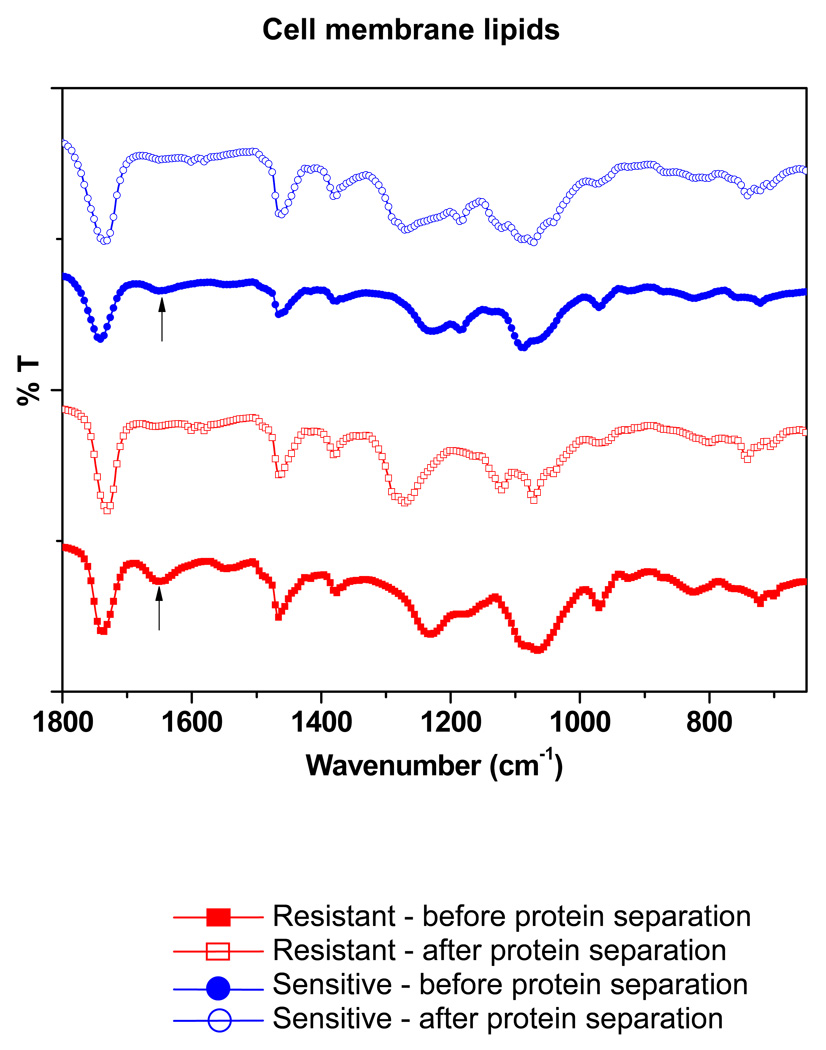
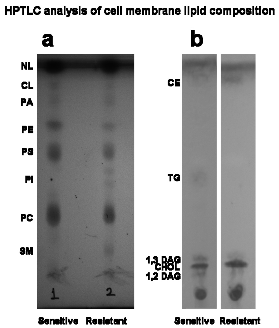
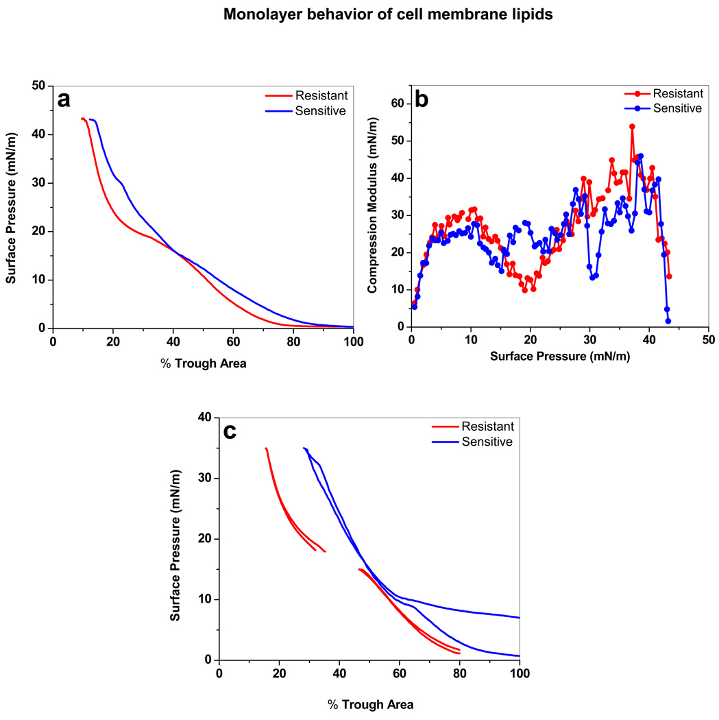
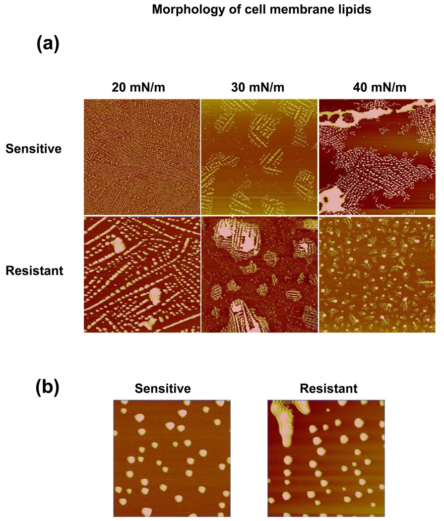
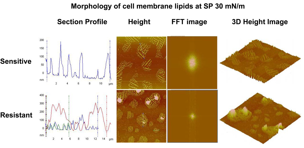
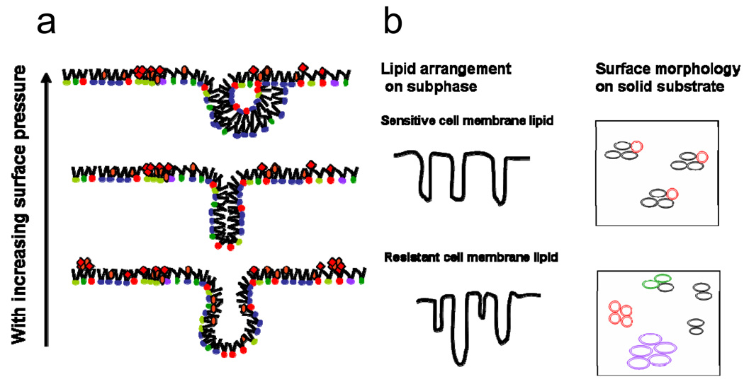
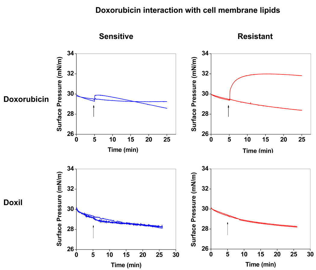
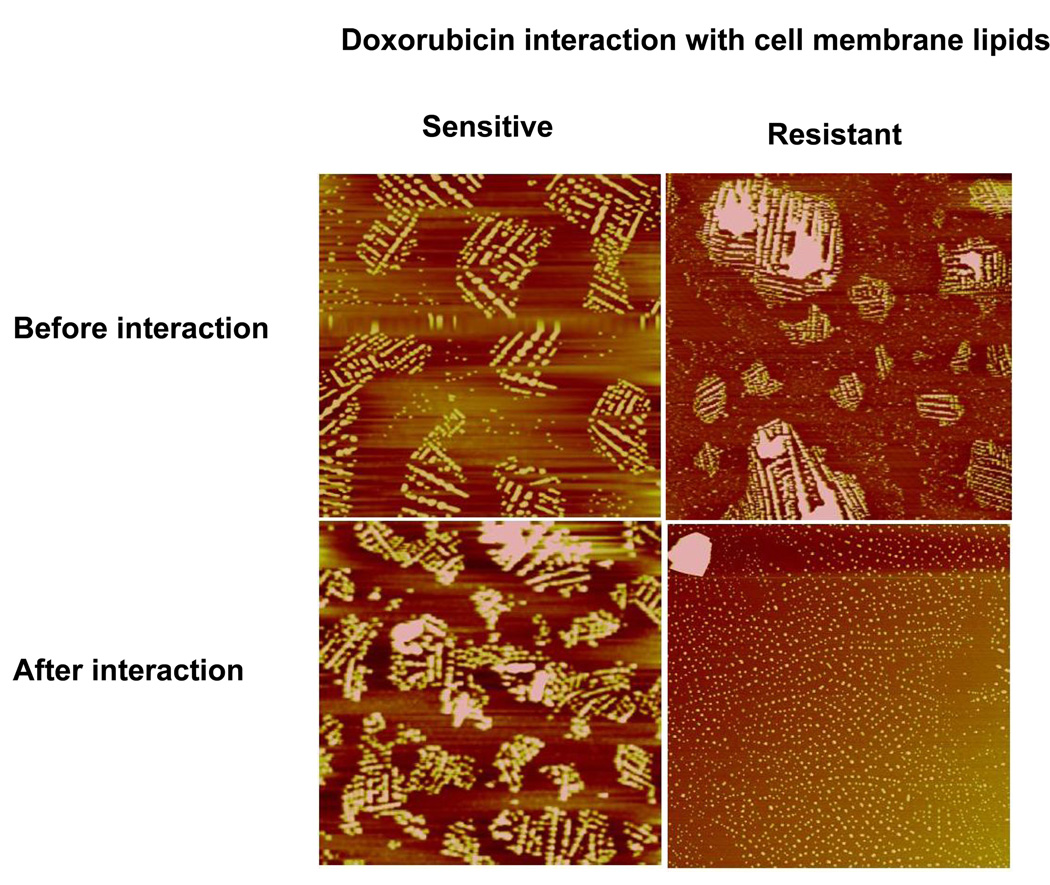
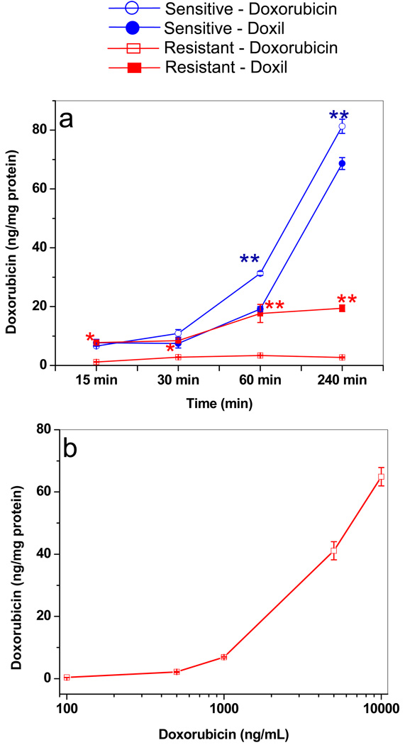
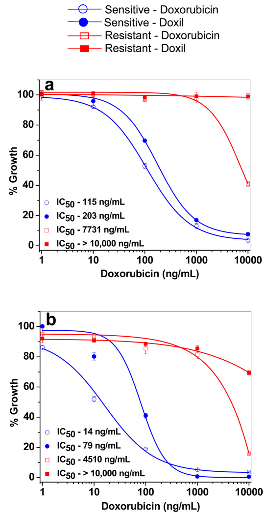
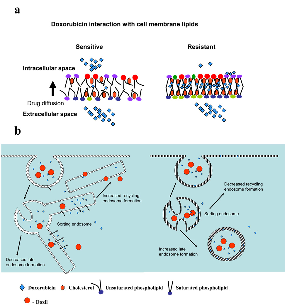
Understanding the role of lipids in drug transport is critical in cancer chemotherapy to overcome drug resistance. In this study, we isolated lipids from doxorubicin-sensitive (MCF-7) and -resistant (MCF-7/ADR) breast cancer cells to characterize the biophysical properties of membrane lipids (particularly lipid packing and membrane fluidity) and to understand the role of the interaction of cell membrane lipids with drug/nanocarrier on drug uptake and efficacy. Resistant cell membrane lipids showed significantly different composition and formed more condensed, less fluid monolayers than did lipids from sensitive cells. Doxorubicin, used as a model anticancer agent, showed a strong hydrophobic interaction with resistant cell membrane lipids but significantly less interaction, as well as a different pattern of interaction (i.e., ionic), with sensitive ones. The threshold intracellular doxorubicin concentration required to produce an antiproliferative effect was similar for both sensitive and resistant cell lines, suggesting that drug transport is a major barrier in determining drug efficacy in resistant cells. In addition to the biophysical characteristics of resistant cell membrane lipids, lipid-doxorubicin interactions appear to decrease intracellular drug transport via diffusion as the drug is trapped in the lipid bilayer. The rigid nature of resistant cell membranes also seems to influence endosomal functions that inhibit drug uptake when a liposomal formulation of doxorubicin is used. In conclusion, biophysical properties of resistant cell membrane lipids significantly influence drug transport, and hence drug efficacy. A better understanding of the mechanisms of cancer drug resistance is vital to developing more effective therapeutic interventions. In this regard, biophysical interaction studies with cell membrane lipids might be helpful to improve drug transport and efficacy through drug discovery and/or drug delivery approaches by overcoming the lipid barrier in resistant cells.


Figures

Figure 1
Figure 1
Analysis of sensitive vs. resistant cell membrane lipids following hydrophobic protein separation. FTIR spectra show the absence of a peak at 1650 cm−1 (shown with arrows), corresponding to a peptide following column separation of lipids.
Figure 2
Figure 2
Phospholipid separation and quantification of different lipids in sensitive vs. resistant cell membrane lipid extracts by HPTLC. Representative data from four different lipid extracts from each cell line.
Figure 3
Figure 3
Biophysical characterization of the lipids isolated from resistant vs. sensitive cells. (a) Compression isotherm (π-A) of sensitive and resistant cell membrane lipids. To obtain a complete isotherm, the lipid extract was spread onto the subphase at different initial SPs, and the lipids were compressed at 5 mm/min. Different parts of the isotherms were collected in two experiments. Data from these experiments were copied into one plot, and the overlapping regions were merged to show the complete isotherm. (b) Compression modulus of Langmuir films over the entire SP range for sensitive vs. resistant cell membrane lipids. Compression modulus was calculated from π-A isotherm data using Cs−1 = − A* (dπ/dA). (c) Compression-expansion isotherm of sensitive vs. resistant cell membrane lipids shows mixing and demixing of lipids.
Figure 4
Figure 4
Morphological analysis of the LB films of resistant vs. sensitive membrane lipids using an AFM. (a) Height images of the LB films of resistant and sensitive cell membrane lipids transferred at different SPs. Scan size = 40 µm; height scale = 300 nm. Resistant and sensitive cell membrane lipids show phase separation but form distinctly different domains upon compression. (b) Magnified AFM images of LB films at SP 20 mN/m. Scan size = 10 µm; height scale = 100 nm.
Figure 5
Figure 5
Morphological analysis of domains of lipids obtained from resistant and sensitive cells. The LB films were transferred at SP 30 mN/m and analyzed using an AFM. Section profiles of the height images show large and more heterogeneous domains for resistant cell membrane lipids than for sensitive cell membrane lipids. FFT images of the corresponding height images revealed that resistant cell membrane lipids form more condensed film than sensitive cell membrane lipids. The 3D height image clearly shows a greater degree of lateral heterogeneity in the LB films of resistant cell membrane lipids than sensitive cell membrane lipids, thus confirming the section profile analysis.
Figure 6
Figure 6
A schematic representation of the monolayer behavior of a complex lipid mixture with compression. (a) Upon lateral compression, saturated and unsaturated phospholipids as well as NLs (cholesterol, triglycerides) become unstable at very high surface density and form bilayer folds. The bilayer folds can bend to form semi-vesicles. (b) A schematic representation of differences in lipid arrangement on the subphase and on solid substrate between sensitive and resistant cell membrane lipids. Lipids extracted from sensitive cells form vesicles, which are uniform and have similar interfacial characteristics, whereas in resistant cells, because of the higher number of lipid species, vesicles with different interfacial characteristics are formed. Domains are formed due to differences in interfacial characteristics, which arise due to differences in the head group, a hydrophobic chain length of lipids in different vesicles.
Figure 7
Figure 7
Changes in SP of sensitive and resistant membrane lipids following interaction with doxorubicin in solution and Doxil. Ten microliters of doxorubicin (1 mg/mL in ethanol:water [1:1 v/v] or 50 µL of Doxil (1 mg/mL doxorubicin) was injected into the subphase, and the change in SP was recorded immediately with time. Similarly, 10 µL of ethanol:water (1:1 v/v) without drug was injected as a control for doxorubicin, whereas 50 µL of water was injected as a control for Doxil. Representative data from four different repeats.
Figure 8
Figure 8
Effect of doxorubicin interaction on the morphology of cell membrane lipids. LB films were transferred at SP 30 mN/m prior to and after interaction with doxorubicin for 20 min. Height scale for all images is 150 nm. The LB film of the sensitive cell membrane lipids did not change following interaction with doxorubicin, but resistant cell membrane lipids showed inhibition of phase separation following interaction with doxorubicin.
Figure 9
Figure 9
Doxorubicin uptake in resistant and sensitive cells. (a) Drug uptake in cells treated with doxorubicin in solution vs. Doxil with incubation time. Uptake increased with longer incubation time in sensitive cells but not in resistant cells. Data as mean ± SEM (n = 4). *p < 0.05 doxorubicin vs. Doxil; **p < 0.005 doxorubicin vs. Doxil. (b) Drug uptake in resistant cells with doxorubicin in solution at different concentrations at 4-h incubation time. Uptake of doxorubicin in resistant cells increased only after ~1,000 ng/mL concentration in culture medium.
Figure 10
Figure 10
Antiproliferative activity of doxorubicin in solution and with Doxil in sensitive and resistant cells following (a) 3-d and (b) 6-d treatment. In 3-d treatment, cells were incubated with different doses of drug, whereas in 6-d treatment, cells were incubated with drug for 3 d and then in drug-free medium for another 3 d prior to MTS assay. Data as mean ± SEM (n = 6). p < 0.005 for IC50 of doxorubicin vs. Doxil in sensitive cells. p < 0.005 for IC50 of doxorubicin or Doxil in sensitive vs. resistant cells.
Figure 11
Figure 11
Mechanism of drug uptake and transport in sensitive and resistant cells (a) Schematic representation of the probable effects of lipid packing density and fluidity of sensitive and resistant cell membrane lipids on drug diffusion across the lipid bilayer. Low lipid packing density and high fluidity arrangement of the lipids of sensitive cells allow the drug to diffuse freely across the bilayer without interaction. In contrast, high lipid packing density and low fluidity of resistant cells and the hydrophobic nature of resistant membrane lipids hinders free drug diffusion and favors drug-lipid interaction, trapping the drug in membrane lipids. (b) Suggested pathways of drug transport in resistant and sensitive cells. Drug or Doxil preferentially follow the pathway of sorting endosomes to recycling endosomes in sensitive cells as against sorting endosomes to late endosomes/lysosomes in resistant cells. This effect could be due to the low membrane fluidity of resistant cells vs. that of sensitive cells and also due the difference in drug interaction with resistant and sensitive cell membrane lipids. Doxorubicin interacts with the membrane lipids of resistant cells but not that of sensitive cells.

Similar articles

Cited by

References

    1. Peetla C, Stine A, Labhasetwar V. Biophysical interactions with model lipid membranes: applications in drug discovery and drug delivery. Mol. Pharmaceutics. 2009;6:1264–1276. - PMC - PubMed
    1. Seddon AM, Casey D, Law RV, Gee A, Templer RH, Ces O. Drug interactions with lipid membranes. Chem. Soc. Rev. 2009;38:2509–2519. - PubMed
    1. Hendrich AB, Michalak K. Lipids as a target for drugs modulating multidrug resistance of cancer cells. Curr. Drug Targets. 2003;4:23–30. - PubMed
    1. Pallarés-Trujillo J, López-Soriano FJ, Argilés JM. Lipids: A key role in multidrug resistance? (Review) Int. J. Oncol. 2000;16:783–798. - PubMed
    1. Pajeva I, Todorov DK, Seydel J. Membrane effects of the antitumor drugs doxorubicin and thaliblastine: comparison to multidrug resistance modulators verapamil and trans-flupentixol. Eur. J. Pharm. Sci. 2004;21:243–250. - PubMed

Publication types