. 2011 Mar;39(4):1351-9.
doi: 10.1093/nar/gkq975. Epub 2010 Oct 18.

Interaction of Rep and DnaB on DNA

Affiliations

Interaction of Rep and DnaB on DNA

John Atkinson et al. Nucleic Acids Res. 2011 Mar.

Abstract

Genome duplication requires not only unwinding of the template but also the displacement of proteins bound to the template, a function performed by replicative helicases located at the fork. However, accessory helicases are also needed since the replicative helicase stalls occasionally at nucleoprotein complexes. In Escherichia coli, the primary and accessory helicases DnaB and Rep translocate along the lagging and leading strand templates, respectively, interact physically and also display cooperativity in the unwinding of model forked DNA substrates. We demonstrate here that this cooperativity is displayed only by Rep and not by other tested helicases. ssDNA must be exposed on the leading strand template to elicit this cooperativity, indicating that forks blocked at protein-DNA complexes contain ssDNA ahead of the leading strand polymerase. However, stable Rep-DnaB complexes can form on linear as well as branched DNA, indicating that Rep has the capacity to interact with ssDNA on either the leading or the lagging strand template at forks. Inhibition of Rep binding to the lagging strand template by competition with SSB might therefore be critical in targeting accessory helicases to the leading strand template, indicating an important role for replisome architecture in promoting accessory helicase function at blocked replisomes.


Figures

Figure 1.
Figure 1.
Cooperativity between DnaB and Rep in unwinding of forked DNA structures requires ssDNA on both leading and lagging strand arms. (A) Unwinding of substrates 1, 2, 3 and 4 by the indicated combinations of Rep and DnaB. (B) The fraction of substrates 1–4 unwound by the indicated combinations of Rep and DnaB. The concentration of Rep was 10 nM and the concentration of hexameric DnaB was 10 nM. Shaded circles indicate the positions of the radiolabel whilst the arrow indicates the 3′ end of oligonucleotides.
Figure 2.
Figure 2.
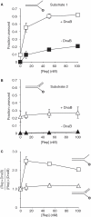
Cooperativity in unwinding by DnaB and Rep is observed on forked DNA bearing a ssDNA leading strand arm but not a dsDNA leading strand arm. (A) and (B) Unwinding of substrates 1 and 2 by the indicated concentrations of Rep in the absence and in the presence of 10 nM DnaB hexamers. (C) Relative levels of substrate unwinding by Rep plus DnaB in comparison to the sum of unwinding by each individual helicase.
Figure 3.
Figure 3.
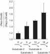
Cooperativity between DnaB and Rep increases with the size of ssDNA exposed on the leading strand template at forks. Unwinding of substrates 2 and 5–7 was monitored in the presence of 10 nM Rep, 10 nM DnaB hexamers and 10 nM Rep +10 nM DnaB hexamers. The relative levels of unwinding by Rep plus DnaB in comparison to the sum of unwinding by each individual helicase is shown.
Figure 4.
Figure 4.
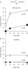
Binding of Rep is not sufficient to enhance DNA unwinding by DnaB. (A) Unwinding of substrate 1 by the indicated concentrations of wild-type Rep in the absence and in the presence of 10 nM DnaB hexamers. (B) Unwinding of substrate 1 by RepK28A without and with 10 nM DnaB hexamers.
Figure 5.
Figure 5.
Cooperativity with DnaB is specific to Rep. Unwinding of substrate 1 by 0, 10, 50 and 100 nM E. coli Rep and DinG, B. stearothermophilus PcrA or D. radiodurans RecD2 was monitored in the absence and in the presence of 10 nM E. coli DnaB hexamers. Relative levels of substrate unwinding by the test helicase plus DnaB in comparison to the sum of unwinding by the individual test helicase and DnaB is shown as a function of the concentration of the test helicase.
Figure 6.
Figure 6.
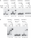
Formation of stable Rep–DnaB–DNA complexes does not require branched DNA. (A) gel mobility shift assays with substrates 1–4 in the presence of 10 nM Rep and 10 nM DnaB hexamers as indicated. (B) gel mobility shift assays of substrates 1, 8 and 9 with 10 nM Rep and 100 nM DnaB hexamers. Note that similar patterns were also observed with substrates 1, 8 and 9 with 10 nM Rep and 10 nM DnaB hexamers (data not shown).
Figure 7.
Figure 7.
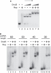
Rep and DnaB can form stable complexes with linear ssDNA. (A) binding of dT60 by 10 nM Rep (lanes 2, 6–8) in the presence of 10, 25 and 100 nM DnaB hexamers as indicated. (B) binding of dT60, dT50, dT40 and dT30 by 10 nM Rep and 100 nM DnaB hexamers as indicated.

References

    1. Mirkin EV, Mirkin SM. Replication fork stalling at natural impediments. Microbiol. Mol. Biol. Rev. 2007;71:13–35. - PMC - PubMed
    1. French S. Consequences of replication fork movement through transcription units in vivo. Science. 1992;258:1362–1365. - PubMed
    1. Deshpande AM, Newlon CS. DNA replication fork pause sites dependent on transcription. Science. 1996;272:1030–1033. - PubMed
    1. McGlynn P, Lloyd RG. Modulation of RNA polymerase by (p)ppGpp reveals a RecG-dependent mechanism for replication fork progression. Cell. 2000;101:35–45. - PubMed
    1. Trautinger BW, Jaktaji RP, Rusakova E, Lloyd RG. RNA polymerase modulators and DNA repair activities resolve conflicts between DNA replication and transcription. Mol. Cell. 2005;19:247–258. - PubMed

Publication types

MeSH terms