. 2010 Dec 31;285(53):41781-94.
doi: 10.1074/jbc.M110.140889. Epub 2010 Oct 19.

CD300 heterocomplexes, a new and family-restricted mechanism for myeloid cell signaling regulation

Affiliations

CD300 heterocomplexes, a new and family-restricted mechanism for myeloid cell signaling regulation

Agueda Martínez-Barriocanal et al. J Biol Chem. .

Abstract

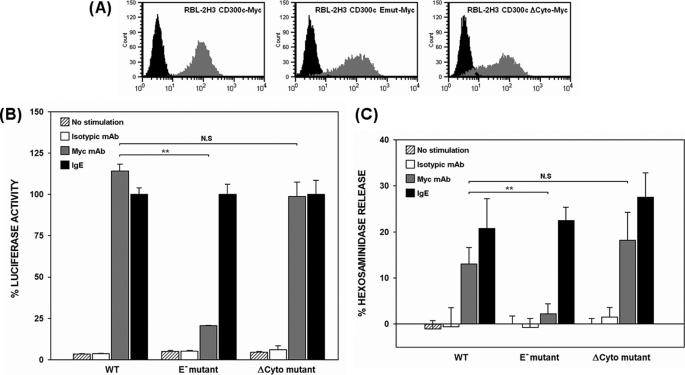
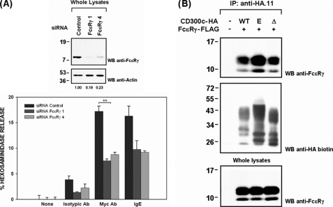
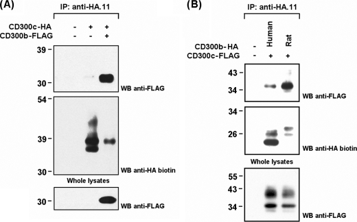
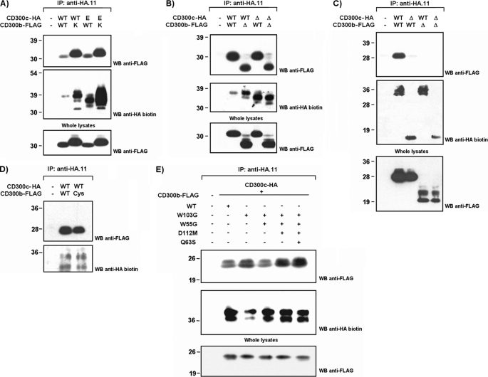
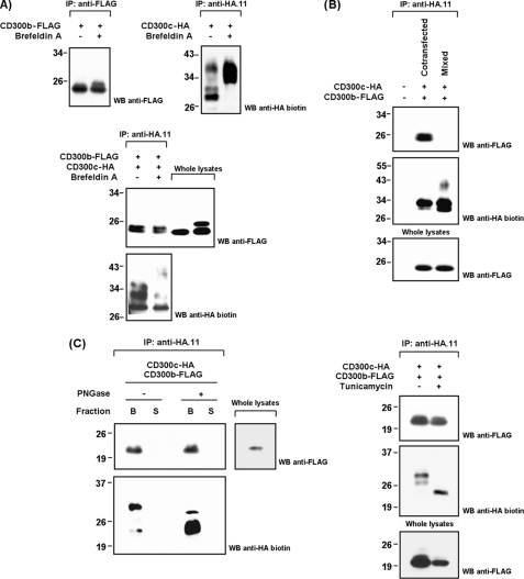
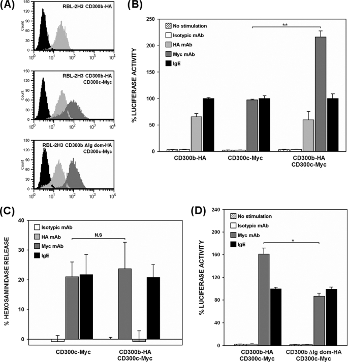
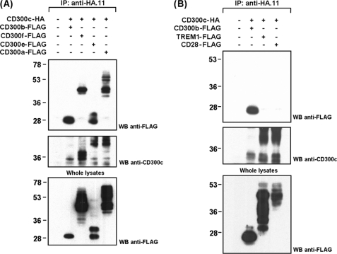
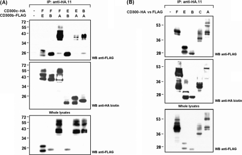
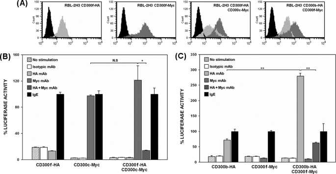
The CD300 family of myeloid immunoglobulin receptors includes activating (CD300b, CD300e) and inhibitory members (CD300a, CD300f), as well as molecules of uncertain function presenting a negative charge within their transmembrane domain (CD300c, CD300d). In this paper, we establish that CD300c is a functional immune receptor able to deliver activating signals upon ligation in RBL-2H3 mast cells. CD300c signaling is partially mediated by a direct association with the immune receptor tyrosine-based activation motif-bearing adaptor FcεRγ. The existence of complementary transmembrane-charged residues in certain CD300 receptors suggested the formation of heterodimers within this family. Indeed, we proved the interaction between CD300b and CD300c in transfected COS-7 cells and demonstrated that it has important functional consequences. Unexpectedly, dimmer formation was dependent on the immunoglobulin domains rather than the charged transmembrane residues. Concordantly, all CD300 members were found to interact with each other, even with themselves, forming both homo- and heterodimers. We found that the combination of CD300 receptors in a complex differentially modulates the signaling outcome, strongly suggesting a new mechanism by which CD300 complexes could regulate the activation of myeloid cells upon interaction with their natural ligands.


Figures

FIGURE 1.
FIGURE 1.
CD300c delivers activating signals in RBL-2H3 cells. A, RBL-2H3 cells were stably transfected with 2×Myc-tagged forms of CD300c. Cell surface expression of CD300c molecules was checked by flow cytometry using anti-Myc9E10 mAb (dark gray histogram) or an isotypic mAb as a negative control (black histogram). B, RBL-2H3 transfectants were transiently transfected with 3×NFAT/AP1-Luciferase and TK-Renilla plasmids. Luciferase activity was measured after stimulation for 7 h with the indicated antibodies. Data were normalized and expressed as a percentage of luciferase activity considering IgE stimulation as the top threshold of activation. Duplicates were performed for all the stimulations. The result is representative of three independent experiments. C, RBL-2H3 transfectants were stimulated with the indicated antibodies to induce cell degranulation. Percentage of β-hexosaminidase release was assessed by incubating the supernatant with 4-nitrophenyl N-acetyl-β-d-glucosaminide substrate. Each assay was set up in triplicate. The result is a mean of three independent experiments (**, p ≤ 0.01). N.S., non-significant. Error bars represent standard deviation.
FIGURE 2.
FIGURE 2.
CD300c signals through FcϵRγ adaptor molecule. A, RBL-2H3 CD300c-2×Myc cells were transiently transfected with control or FcϵRγ siRNAs. Forty-eight hours after transfection a subset of cells were lysed and proteins (2 μg) were run on 15% SDS-PAGE and transferred to PVDF filters. FcϵRγ interference was assessed by Western blot (WB). Values correspond to FcϵRγ expression, which was calculated by quantifying the intensity of FcϵRγ versus actin. Hexosaminidase release was assessed as described. Triplicates were performed for all the stimulations. The result is representative of two independent experiments. B, COS-7 cells were transiently transfected with HA-tagged CD300c WT or mutant forms (E191V and ΔCyto) in combination with FcϵRγ-FLAG. Cell lysates (0.5% CHAPS) were immunoprecipitated with anti-HA.11 mAb and analyzed by 15% SDS-PAGE under reducing conditions. Proteins were transferred to PVDF filter and probed with the indicated antibodies. Whole cell lysates (2%) were included as controls (**, p ≤ 0.01). Error bars represent standard deviation.
FIGURE 3.
FIGURE 3.
CD300c associates with both human and rat CD300b in transfected COS-7 cells. A, COS-7 cells were transiently transfected with HA-tagged CD300c alone or in combination with FLAG-tagged CD300b. Cell were lysed (1% Triton X-100) and immunoprecipitated (IP) with anti-HA.11 mAb. Blots were probed with the indicated antibodies. Whole cell lysates (2%) were included as controls. B, same experiment as in A to compare the recruitment of CD300b-FLAG from human and rat origin by CD300c-HA. WB, Western blot.
FIGURE 4.
FIGURE 4.
CD300c and CD300b interact through their Ig-like domains. COS-7 cells were transiently transfected with HA-tagged CD300c in combination with FLAG-tagged CD300b. Wild type forms were tested against transmembrane substitution mutants (A), intracellular deletion mutants (B), extracellular deletion mutants (C), cysteine substitution mutants (D), or point mutants affecting the protruding body in the Ig fold (E) to map the interaction between both molecules. Cells were lysed (1% Triton X-100) and CD300c was immunoprecipitated (IP) with anti-HA.11 mAb in all cases. Western blots (WB) were conducted with the indicated Abs. Whole cell lysates (2%) were included as controls.
FIGURE 5.
FIGURE 5.
CD300c and CD300b form cis-interacting complexes in the ER. A, COS-7 cells were transiently transfected with CD300 constructions. Forty-eight hours post-transfection cells were stimulated for 24 h with brefeldin A (1 μg/ml) or vehicle (70% EtOH). Cells were lysed (1% Triton X-100) and immunoprecipitated (IP) with the indicated antibodies. Western blots (WB) were conducted as described. Whole cell lysates (2%) were included as controls when assessing co-transfections. B, CD300c-HA and CD300b-FLAG were expressed both individually and simultaneously in COS-7. When expressed individually, clarificates where processed independently until the immunoprecipitation step with anti-HA.11 mAb. Western blots were conducted normally. C, immunoprecipitates from COS-7 co-transfected cells were subjected to peptide:N-glycosidase treatment in non-denaturing conditions. The eluted and immobilized fractions were analyzed separately (S = supernatant and B = beads) (left panel). COS-7 cells were transiently cotransfected with CD300 constructions. Eighteen hours post-transfection cells were stimulated for 48 h with tunicamycin (1 μg/ml) or vehicle (dimethyl sulfoxide). Cells were lysed and immunoprecipitated as described above (right panel).
FIGURE 6.
FIGURE 6.
CD300b acts as a modifier of CD300c signaling. A, RBL-2H3 cells stably expressing CD300c-Myc on the cell surface were transfected with CD300b-HA or CD300b ΔIg-HA. Cell surface expression of the different transfectants was checked by flow cytometry using anti-Myc9E10 for CD300c (light gray histogram) and anti-HA12CA5 mAb for CD300b (dark gray histogram). Isotypic mAb was used as negative control (black histogram). B and D, RBL-2H3 transfectants were transiently transfected with 3×NFAT/AP1-Luciferase and TK-Renilla plasmids. Luciferase activity was measured after stimulation for 7 h with the indicated antibodies. Data were normalized as described before. Duplicates were performed for all stimulations. Results are representative of five independent experiments. C, RBL-2H3 transfectants were stimulated with the indicated Abs for assessing hexosaminidase granule release. Data are expressed as a percentage of specific release. Each stimulation point was set up in triplicate. The result is a mean of three independent experiments (*, p ≤ 0.05; **, p ≤ 0.01). N.S., non-significant. Error bars represent standard deviation.
FIGURE 7.
FIGURE 7.
CD300c interacts with all CD300 family members but not with other Ig-like receptors. CD300c-HA was transiently transfected in COS-7 cells in combination with FLAG-tagged CD300 receptors (CD300a, CD300b, CD300e, and CD300f) (A) or CD300-related Ig-like receptors (TREM-1 and CD28) (B). Cell lysates (1% Triton X-100) were immunoprecipitated (IP) with anti-HA.11 mAb. Filters were probed with the indicated antibodies. Whole cell lysates (2%) were included as controls. WB, Western blot.
FIGURE 8.
FIGURE 8.
CD300 hetero- and homodimerization. CD300a, CD300b, CD300c, CD300e, and CD300f (HA- or FLAG-tagged) were transiently cotransfected in COS-7 cells following all possible combinations for the detection of hetero- (A) and homodimers (B). Immunoprecipitations (IP) were performed against HA-tagged receptors with anti-HA.11 mAb after cell lysis (1% Triton X-100). Filters were proved with the indicated antibodies. Whole cell lysates (2%) were included as controls. WB, Western blot.
FIGURE 9.
FIGURE 9.
CD300 functional behavior is modified based on complex composition. A, CD300 receptors (Myc or HA-tagged) stably transfected on RBL-2H3 cell surface were assessed by flow cytometry using anti-Myc9E10 (light gray histograms) and anti-HA12CA5 mAb (dark gray histograms). Isotypic mAb was used as negative control (black histograms). B and C, RBL-2H3 transfectants were transiently transfected with 3×NFAT/AP1-Luciferase and TK-Renilla plasmids. Luciferase activity was measured after stimulation for 7 h with the indicated antibodies. Data are normalized as described before. Duplicates were performed for all stimulations. Results are representative of three and two independent experiments respectively (*, p ≤ 0.05; **, p ≤ 0.01). N.S., non-significant. Error bars represent standard deviation.

References

    1. Veillette A., Latour S., Davidson D. (2002) Annu. Rev. Immunol. 20, 669–707 - PubMed
    1. Long E. O. (2008) Immunol. Rev. 224, 70–84 - PMC - PubMed
    1. Tomasello E., Bléry M., Vély F., Vivier E. (2000) Semin. Immunol. 12, 139–147 - PubMed
    1. Humphrey M. B., Lanier L. L., Nakamura M. C. (2005) Immunol. Rev. 208, 50–65 - PubMed
    1. Underhill D. M., Goodridge H. S. (2007) Trends Immunol. 28, 66–73 - PubMed

Publication types

LinkOut - more resources