. 2011 Jan 27;117(4):1400-7.
doi: 10.1182/blood-2010-05-287623. Epub 2010 Oct 19.

von Willebrand factor-mediated platelet adhesion is critical for deep vein thrombosis in mouse models

Affiliations

von Willebrand factor-mediated platelet adhesion is critical for deep vein thrombosis in mouse models

Alexander Brill et al. Blood. .

Abstract

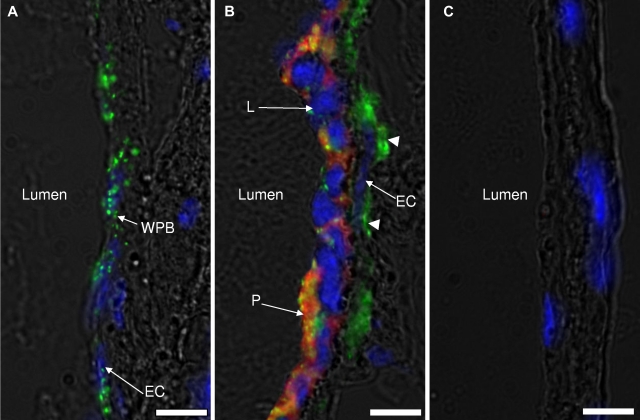
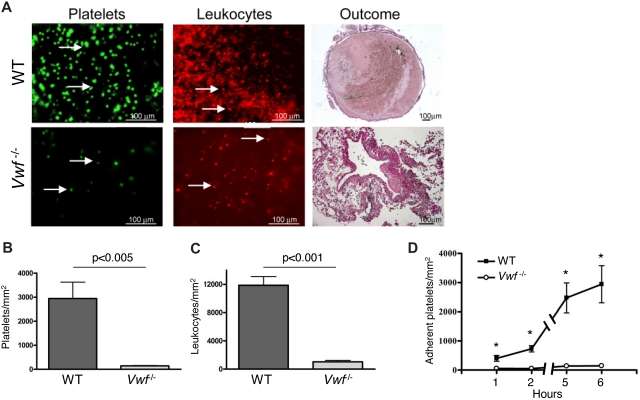
Deep vein thrombosis (DVT) and its complication, pulmonary embolism, are frequent causes of disability and mortality. Although blood flow disturbance is considered an important triggering factor, the mechanism of DVT initiation remains elusive. Here we show that 48-hour flow restriction in the inferior vena cava (IVC) results in the development of thrombi structurally similar to human deep vein thrombi. von Willebrand factor (VWF)-deficient mice were protected from thrombosis induced by complete (stasis) or partial (stenosis) flow restriction in the IVC. Mice with half normal VWF levels were also protected in the stenosis model. Besides promoting platelet adhesion, VWF carries Factor VIII. Repeated infusions of recombinant Factor VIII did not rescue thrombosis in VWF(-/-) mice, indicating that impaired coagulation was not the primary reason for the absence of DVT in VWF(-/-) mice. Infusion of GPG-290, a mutant glycoprotein Ibα-immunoglobulin chimera that specifically inhibits interaction of the VWF A1 domain with platelets, prevented thrombosis in wild-type mice. Intravital microscopy showed that platelet and leukocyte recruitment in the early stages of DVT was dramatically higher in wild-type than in VWF(-/-) IVC. Our results demonstrate a pathogenetic role for VWF-platelet interaction in flow disturbance-induced venous thrombosis.


Figures

Figure 1
Figure 1
VWF−/− mice are protected from flow restriction–induced venous thrombosis. IVC stenosis (A-C) or stasis (D-F) in WT, VWF+/−, and VWF−/− mice was produced. After 48 hours, mice were killed and thrombi were harvested. Values for weight and length of the thrombi are presented in A-B (stenosis model) and D-E (stasis model). The percentage of mice with a thrombus is shown in panels C and F for stenosis and stasis models, respectively. Horizontal bars in dot plots represent median. Stenosis model: WT, n = 6; VWF+/−, n = 10; VWF−/−, n = 14. Stasis model: WT, n = 11; VWF+/−, n = 10; VWF−/−, n = 9.
Figure 2
Figure 2
IVC stenosis induces formation of human-like deep vein thrombi in WT mice. Stenosis of IVC was applied in WT mice, and a typical thrombus formed in 48 hours is presented (A). “White” (W) and “red” (R) regions of the thrombus are indicated. Bar, 500 μm. (B-C) A thrombus obtained in the stenosis model was longitudinally cut and stained for a platelet marker (αIIb, green), fibrinogen/fibrin (red), and counterstained for nuclei (blue). Combined images of the thrombus regions remote from (B) and close to (C) the ligation is shown. Unstained dark area is rich in red blood cells. Bar, 50 μm. Representative photographs are shown, n = 3.
Figure 3
Figure 3
GPG-290 prevents thrombus development in the stenosis model without affecting leukocyte rolling. Stenosis of IVC was performed in WT mice. GPG-290 (100 μg/kg) or sterile saline was infused immediately after operation and again after 24 hours. Thrombi were harvested 48 hours after operation. Values for weight (A) and length (B) of the thrombi are shown with medians (horizontal bars). (C) Incidence of the thrombosis. Vehicle, n = 9; GPG-290, n = 11. (D) Leukocyte rolling was recorded in the same mesenteric vein (diameter 200-300 μm) before and after infusion of GPG-290 (5 mg/kg). No effect of GPG-290 on the number of rolling leukocytes was observed. Results are mean ± SEM, n = 4. (E) Representative photographs of rolling leukocytes in a mesenteric vein before and after infusion of GPG-290. White arrow indicates a rolling leukocyte. Bar, 50 μm.
Figure 4
Figure 4
Weibel-Palade body release in IVC endothelium after flow restriction. IVCs from WT mice 6 hours after stenosis had been applied were rapidly excised and snap-frozen in Optimal Cutting Temperature compound (Tissue-Tek). The part of IVC within 2 mm from the stenosis site was sectioned, immunostained for VWF (green), platelet αIIb (red), counterstained for nuclei (blue), and photographed by an inverted fluorescent microscope. Two types of endothelial zones were observed after 6 hours of stenosis: (A) a nonactivated endothelial zone with the typical WPB (green, WPBs) staining pattern and (B) a zone of activated endothelium with no obvious WPB staining. Leukocytes (L) and platelets (P) are adherent to the endothelial cell layer (EC) only in panel B. Subendothelially located VWF is designated with arrowheads. (C) Control staining with nonspecific IgG used instead of primary antibodies. Representative images of 3 mice are shown. Bar, 10 μm.
Figure 5
Figure 5
Platelet and leukocyte recruitment is impaired in VWF−/− mice. (A) IVC stenosis was produced in WT (top row) and VWF−/− (bottom row) mice. After 6 hours, fluorescently labeled platelets or Rhodamine 6G were intravenously infused, and the cell accumulation at the area 1-2 mm below the IVC suture (toward the tail) was examined by intravital microscopy using a fluorescent microscope at ×20 magnification. Outcome: thrombus formed within 48 hours after IVC stenosis induction in a WT but not a VWF−/− mouse. (B-C) Quantification of adherent platelets and leukocytes, respectively, 6 hours after flow restriction. (D) Kinetics of platelet accumulation 1-6 hours after flow restriction in the IVC in WT and VWF−/− mice. Data are shown as mean ± standard error of the mean. *Statistically significant difference between WT and VWF−/− mice. WT, n = 8-10; VWF−/−, n = 3.

Comment in

  • Clues to DVT pathogenesis.
    Motto D. Motto D. Blood. 2011 Jan 27;117(4):1106-7. doi: 10.1182/blood-2010-11-315879. Blood. 2011. PMID: 21273329

References

    1. Heit JA. The epidemiology of venous thromboembolism in the community. Arterioscler Thromb Vasc Biol. 2008;28(3):370–372. - PMC - PubMed
    1. Sevitt S. The structure and growth of valve-pocket thrombi in femoral veins. J Clin Pathol. 1974;27(7):517–528. - PMC - PubMed
    1. Esmon CT. Basic mechanisms and pathogenesis of venous thrombosis. Blood Rev. 2009;23(5):225–229. - PMC - PubMed
    1. Poort SR, Rosendaal FR, Reitsma PH, Bertina RM. A common genetic variation in the 3′-untranslated region of the prothrombin gene is associated with elevated plasma prothrombin levels and an increase in venous thrombosis. Blood. 1996;88(10):3698–3703. - PubMed
    1. Griffin JH, Evatt B, Zimmerman TS, Kleiss AJ, Wideman C. Deficiency of protein C in congenital thrombotic disease. J Clin Invest. 1981;68(5):1370–1373. - PMC - PubMed

Publication types

MeSH terms