. 2011 Jan 13;117(2):591-4.
doi: 10.1182/blood-2010-03-275305. Epub 2010 Oct 19.

CAL-101, a p110delta selective phosphatidylinositol-3-kinase inhibitor for the treatment of B-cell malignancies, inhibits PI3K signaling and cellular viability

Affiliations

CAL-101, a p110delta selective phosphatidylinositol-3-kinase inhibitor for the treatment of B-cell malignancies, inhibits PI3K signaling and cellular viability

Brian J Lannutti et al. Blood. .

Abstract

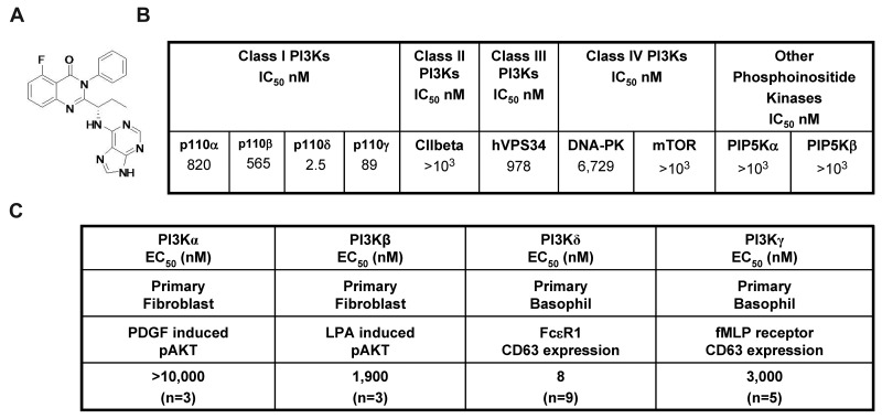
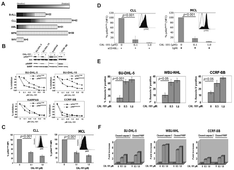
Phosphatidylinositol-3-kinase p110δ serves as a central integration point for signaling from cell surface receptors known to promote malignant B-cell proliferation and survival. This provides a rationale for the development of small molecule inhibitors that selectively target p110δ as a treatment approach for patients with B-cell malignancies. We thus identified 5-fluoro-3-phenyl-2-[(S)-1-(9H-purin-6-ylamino)-propyl]-3H-quinazolin-4-one (CAL-101), a highly selective and potent p110δ small molecule inhibitor (half-maximal effective concentration [EC(50)] = 8nM). Using tumor cell lines and primary patient samples representing multiple B-cell malignancies, we have demonstrated that constitutive phosphatidylinositol-3-kinase pathway activation is p110δ-dependent. CAL-101 blocked constitutive phosphatidylinositol-3-kinase signaling, resulting in decreased phosphorylation of Akt and other downstream effectors, an increase in poly(ADP-ribose) polymerase and caspase cleavage and an induction of apoptosis. These effects have been observed across a broad range of immature and mature B-cell malignancies, thereby providing a rationale for the ongoing clinical evaluation of CAL-101.


Figures

Figure 1
Figure 1
CAL-101 inhibits PI3Kδ with high selectivity. (A) Chemical structure of CAL-101. (B) CAL-101 in vitro activity profiles (half-maximal inhibitory concentration values) against recombinant enzymes of class I, II, III, and IV PI3Ks. CAL-101 was diluted in dimethyl sulfoxide at a concentration of 10mM, and 10-point kinase inhibitory activities were measured over a concentration range (5.0-104nM) with adenosine triphosphate at a concentration consistent with each enzymes Kms. (C) Potency of CAL-101 in PI3K class I isoform-specific cell-based assays. For the analysis of p110α and p110β signaling, murine embryo fibroblasts were stimulated with PDGF or LPA and soluble protein was analyzed by Western blotting for Akt and pAkt473 levels. For the analysis of p110δ and p110γ signaling, basophil activation was measured in isolated peripheral blood mononuclear cell or whole blood using the Flow2 CAST kit according to the manufacturer's standardized methods (Buhlman Laboratories AG). p110δ was activated with anti-FCϵRI, and p110γ was activated with formyl-methionyl-leucyl-phenylalanine. To monitor the basophil cell population and cellular activation, anti–CD63-FITC and anti–CCR3-phycoerythrin antibodies were added to each sample. Cells were fixed and analyzed on a FC500MPL flow cytometer (Beckman Coulter).
Figure 2
Figure 2
CAL-101 inhibits PI3K signaling and cellular viability. (A) CAL-101 screening of primary cells from patients with ALL, CLL, acute myeloid leukemia (AML), or myeloproliferative neoplasm (MPN) or from normal healthy volunteers (NHV). Cell viability was determined using an 3-carboxymethoxyphenyl)-2-(4-sulphenyl)-2H-tetrazolium assay, and the sensitivity of each sample relative to untreated cells was calculated for estimation of the EC50. The heat map readout shown was generated using GenePattern Version 3.2 software (Broad Institute) and indicates the percentage EC50 for each sample relative to the maximum drug concentration tested (10μM). (B) CAL-101 inhibition of p110δ blocks PI3K signaling in malignant B-cell lines and primary patient tumor cells. Serum-starved cells were incubated with 1μM CAL-101, and total cell lysates were subjected to Western blot analysis using anti–phospho-AktS473, anti-Akt, anti-phospho S6S235/236, and anti-S6 antibodies (supplemental data). Starved cells were incubated with vehicle or serial dilution of CAL-101 for 1 hour, and pAktT308, pS6S235/236, total Akt, and total S6 were detected by PathScan sandwich ELISA (supplemental data). (C-D) CLL (n = 5) and MCL (n = 5) patient whole blood samples were subjected to Ficoll-Hypaque separation. Isolated cells were incubated in RPMI with vehicle or serial dilutions of CAL-101 before fixation and staining with anti–phospho-AktT308 Alexa Fluor 488 or isotype-matched Alexa Fluor 488 antibody. Cells pretreated with CAL-101 or vehicle for 1 hour and then stimulated with 10 μg/mL anti-IgM (BCR activation), or 50 ng/mL sCD40L (CD40 stimulation) for 10 minutes before fixation and staining with anti–phospho-AktS473 Alexa Fluor 488 or isotype-matched Alexa Fluor 488 antibody. FITC-CD5+ cells were gated and analyzed by 2-color flow cytometry to quantify intracellular p-AktT308 levels using the Beckman Coulter Cytomics FC 500MPL using CXP Version 2.2 software. Bar graphs represent the percentage difference in mean fluorescence intensity values between isotype-matched control Ig and phospho-AktT308. (E) CAL-101 induces apoptosis in diffuse large B-cell lymphoma, follicular lymphoma, and B-ALL cell lines. Cells were treated with vehicle or 0.5μM or 1.0μM CAL-101 for 24 hours. The percentage of apoptotic cells was determined by annexin V–FITC/7-AAD staining followed by 2-color flow cytometric analysis. Percentages represent both annexin V–FITC/7-AAD negative and annexin V–FITC/7-AAD double-positive. (F) Cells were cultured in RPMI/10% fetal bovine serum with CAL-101 or vehicle alone for 24 hours, and cells were lysed and analyzed by PathScan Sandwich 96-well ELISA for the detection of cleaved caspase-3 and cleaved poly(ADP-ribose) polymerase as indicated (supplemental data). Results are expressed as mean ± SD. Statistically significant differences between means were determined using a one-way analysis of variance. Data are expressed as the fold change and are representative of 3 separate experiments.

References

    1. Engelman JA. Targeting PI3K signalling in cancer: opportunities, challenges and limitations. Nat Rev Cancer. 2009;9(8):550–562. - PubMed
    1. Witzig TE, Kaufmann SH. Inhibition of the phosphatidylinositol 3-kinase/mammalian target of rapamycin pathway in hematologic malignancies. Curr Treat Options Oncol. 2006;7(4):285–294. - PubMed
    1. Bernal A, Pastore RD, Asgary Z, et al. Survival of leukemic B cells promoted by engagement of the antigen receptor. Blood. 2001;98(10):3050–3057. - PubMed
    1. Davis RE, Ngo VN, Lenz G, et al. Chronic active B-cell-receptor signalling in diffuse large B-cell lymphoma. Nature. 2010;463(7277):88–92. - PMC - PubMed
    1. Munk Pedersen I, Reed J. Microenvironmental interactions and survival of CLL B-cells. Leuk Lymphoma. 2004;45(12):2365–2372. - PubMed

MeSH terms