Review
. 2010 Oct;90(4):1461-506.
doi: 10.1152/physrev.00057.2009.

The ß subunit of voltage-gated Ca2+ channels

Affiliations
Review

The ß subunit of voltage-gated Ca2+ channels

Zafir Buraei et al. Physiol Rev. 2010 Oct.

Abstract

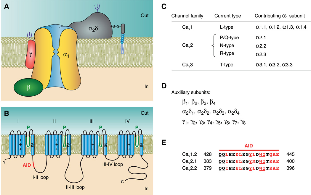
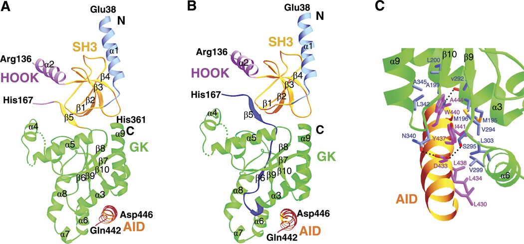
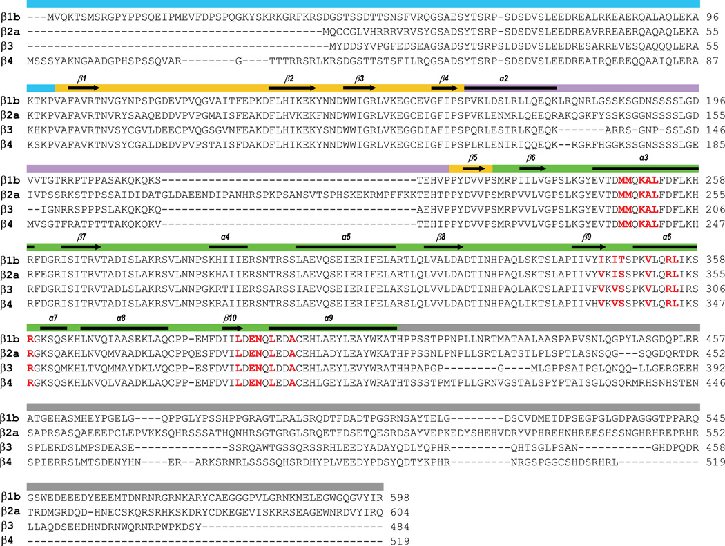
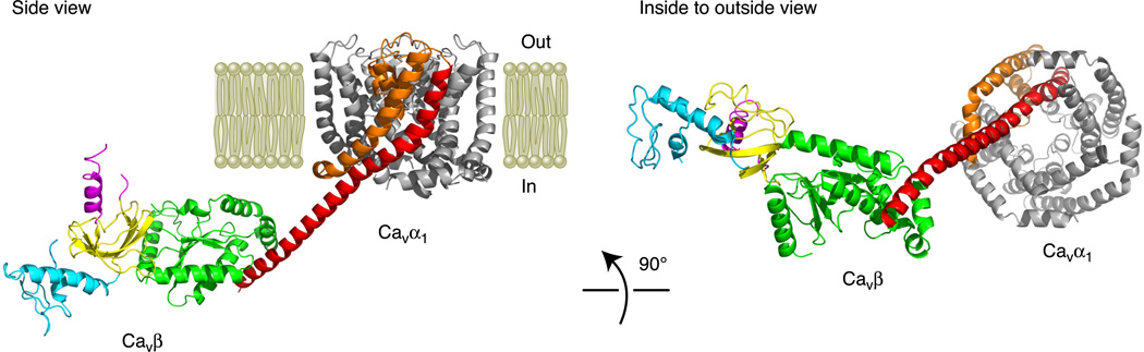
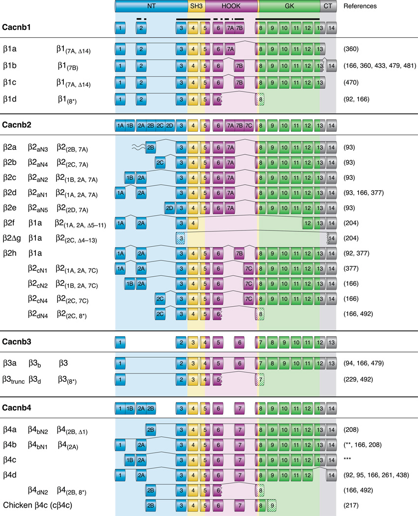
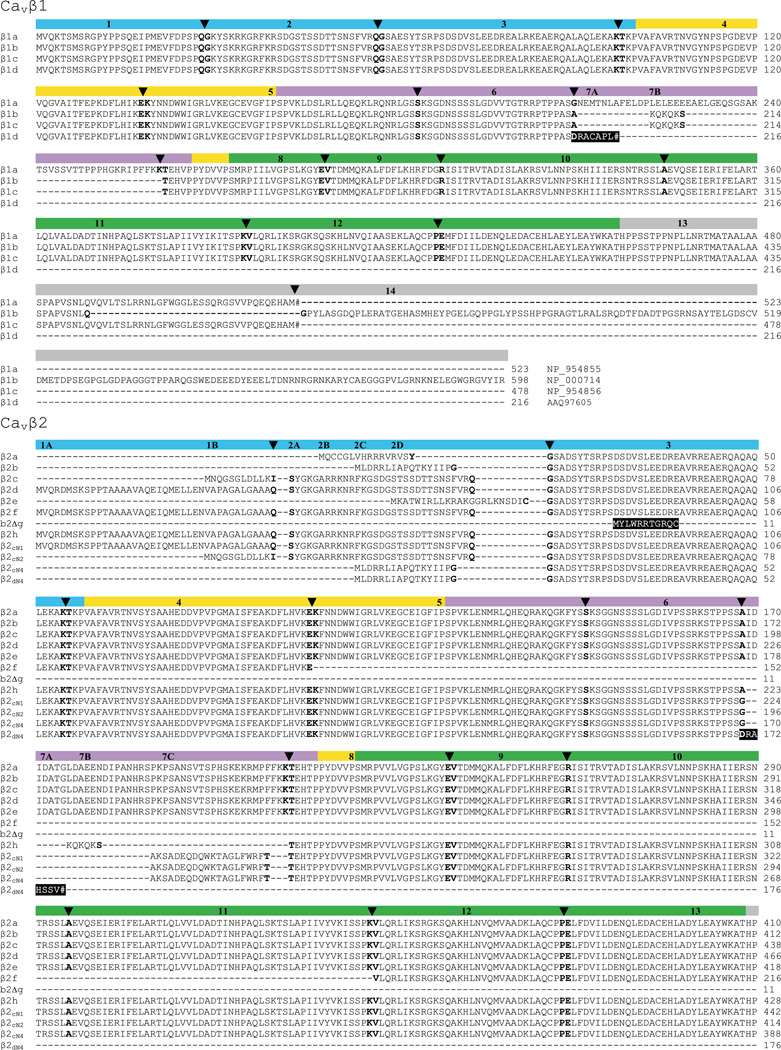
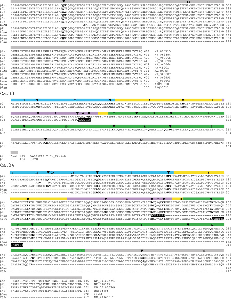
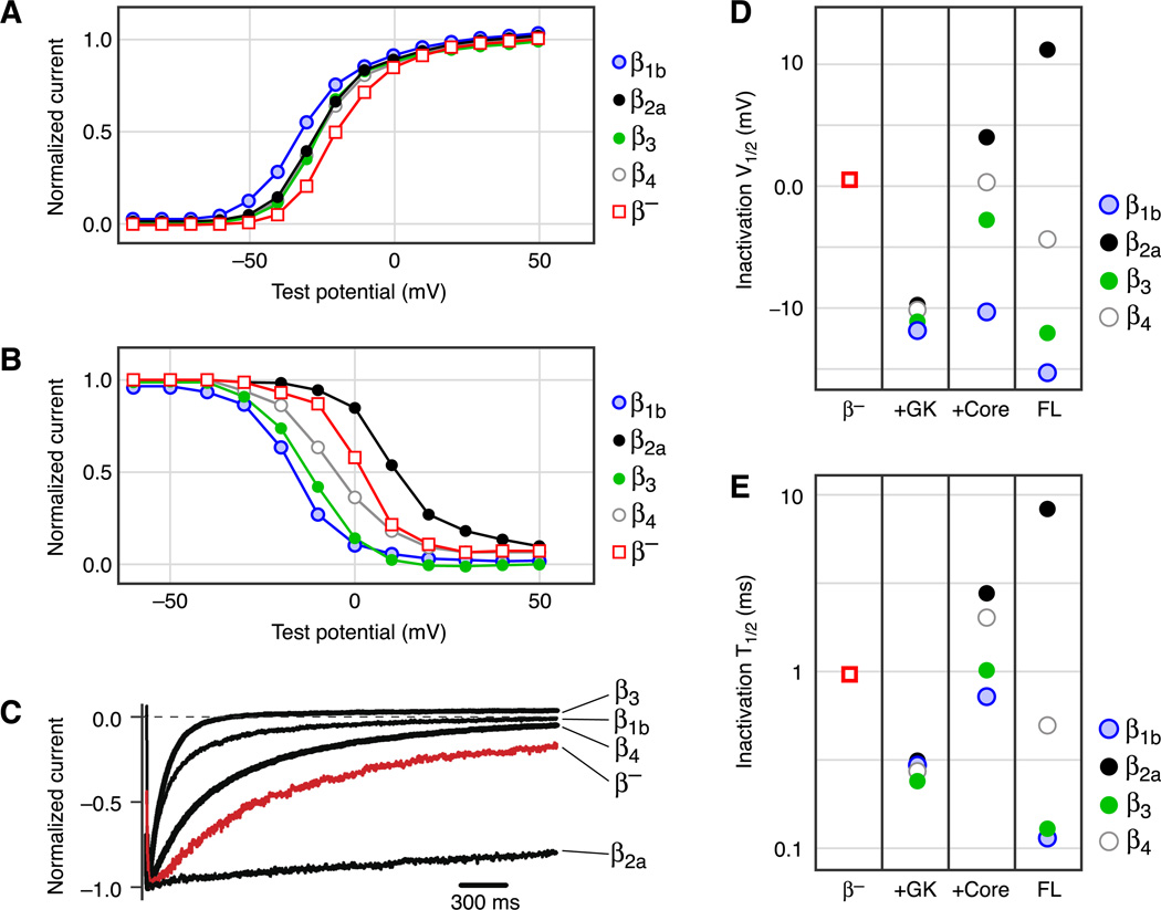
Calcium regulates a wide spectrum of physiological processes such as heartbeat, muscle contraction, neuronal communication, hormone release, cell division, and gene transcription. Major entryways for Ca(2+) in excitable cells are high-voltage activated (HVA) Ca(2+) channels. These are plasma membrane proteins composed of several subunits, including α(1), α(2)δ, β, and γ. Although the principal α(1) subunit (Ca(v)α(1)) contains the channel pore, gating machinery and most drug binding sites, the cytosolic auxiliary β subunit (Ca(v)β) plays an essential role in regulating the surface expression and gating properties of HVA Ca(2+) channels. Ca(v)β is also crucial for the modulation of HVA Ca(2+) channels by G proteins, kinases, and the Ras-related RGK GTPases. New proteins have emerged in recent years that modulate HVA Ca(2+) channels by binding to Ca(v)β. There are also indications that Ca(v)β may carry out Ca(2+) channel-independent functions, including directly regulating gene transcription. All four subtypes of Ca(v)β, encoded by different genes, have a modular organization, consisting of three variable regions, a conserved guanylate kinase (GK) domain, and a conserved Src-homology 3 (SH3) domain, placing them into the membrane-associated guanylate kinase (MAGUK) protein family. Crystal structures of Ca(v)βs reveal how they interact with Ca(v)α(1), open new research avenues, and prompt new inquiries. In this article, we review the structure and various biological functions of Ca(v)β, with both a historical perspective as well as an emphasis on recent advances.


Conflict of interest statement

DISCLOSURES

No conflicts of interest, financial or otherwise, are declared by the authors.

Figures

Fig. 1
Fig. 1
Molecular organization of voltage-gated Ca2+ channels. A: subunit composition of high-voltage activated (HVA) Ca2+ channels. B: schematic representation of the predicted transmembrane topology of Cavα1, with the location of the α-interaction domain (AID) marked. C: Ca2+ channel current types and the corresponding α1 subunits of the channels that produce them. D: list of all cloned auxiliary HVA Ca2+ channel subunits. E: amino acid sequence alignment of the AID from the indicated Cavα1. Residues involved in interactions with Cavβ are marked in red, with the most critical residues underlined. Residue numbers are indicated on both sides of the sequence.
Fig. 2
Fig. 2
Cavβ crystal structure. A: crystal structure of the β3 core in complex with the AID (PDB accession code 1VYT). This structure reveals the following regions: the NH2 terminus (light blue, residues –59), an SH3 domain (gold, residues 60–120 and 170–175), a HOOK region (purple, residues –169), and a GK domain (green, residues 176–360). Residues 137–166 were disordered and are not included. Residues 226–244 (forming the α4 helix of the GK domain) were disordered in this molecule but were well-resolved in another one in the same asymmetric unit. Residues 422–446 of Cav1.2 containing the entire AID are colored in orange. B: same structure as in A but with the BID (β3 residues K163-T193) highlighted in dark blue. The BID spans parts of the SH3-HOOK-GK motif but is not directly involved in binding the AID. C: close-up of the interface between β3 and AID. Some residues involved in the interactions are shown. [Adapted from Chen et al. (84)].
Fig. 3
Fig. 3
Amino acid sequence alignment of Cavβ subtypes. The four included subtypes are β1b (GenBank accession number, NP-000714), β2a (M80545), β3 (M88751), and β4a (L02315). Light blue indicates the NH2 terminus, gold the SH3 domain, purple the HOOK region, green the GK domain, and gray the COOH terminus. Secondary structure elements are indicated in the top line as arrows for β sheets and solid lines for α helices (based on the crystal structure of β3). Residues involved in interactions with the AID are marked in red.
Fig. 4
Fig. 4
Structural model of a partial Cavα1/Cavβ complex on the plasma membrane. A side view and an inside-to-outside view are presented. The partial structure of Cavα1 includes only the S5, P-loop, and S6 segments and is based on a Cavα1 homology model developed in Stary et al. (411). IS5 is colored orange, and IS6 is red. The IS6-AID linker from Cav1.2 is modeled as an α-helix and is joined with IS6 at its NH2 terminus and the AID at its COOH terminus. The structure of Cavβ is based on the crystal structure of the β4 core region (84) and the NMR structure of the β4 NH2 terminus (451); there is no Cavβ COOH terminus. Since the structure of the β4-AID complex is not available, we docked the AID to β4 based on the crystal structure of the β3 core-AID complex (84). The regions of Cavβ are color coded as in Figures 2 and 3 (NH2 terminus in light blue, SH3 in gold, HOOK in purple, and GK in green).
Fig. 5
Fig. 5
Human Cavβ splice variants. Fourteen Cavβ exons (13 for β3) are color-coded based on the regions they give rise to: the NH2 terminus (light blue), the SH3 domain (gold), the HOOK (purple), the GK domain (green), and the COOH terminus (gray). Exons are numbered, and some exons have additional letters to indicate alternatively spliced variants. The thick full and dashed lines at the very top indicate highly or somewhat conserved exons, respectively. Of the weakly conserved regions, similar exons are placed in the same column (e.g., β1 exon 2 is homologous to β2 exon 2A). Exons 13 and 14 of β1 were originally designated as 13a and 13b, respectively (222). The names of splice variants are, from left to right columns, those used in this article, those proposed by Foell et al. (166), and those proposed by Yang and Berggren (486). β2a is the only splice variant that can be palmitoylated (wave). The jagged edge (e.g., exon 6 of β1d) indicates missing amino acids resulting from exon skipping and/or frame-shifts. Striped exons (e.g., exon 8 of β1d) are translated with a frame shift; hence, their amino acid sequence is unrelated to the “conventional” sequence produced by that exon. **Direct submission by M. E. Williams, 1997. ***AK316045; direct submission by T. Isogai and J. Yamamoto, 2008.
Fig. 6
Fig. 6
Amino acid sequence alignment of Cavβ splice variants. The 5 Cavβ regions, their corresponding exons, and the exon boundaries are marked. Color coding follows the same scheme as in previous figures, with the NH2 terminus in light blue, the SH3 domain in gold, the HOOK in purple, the GK domain in green, and the COOH terminus in gray. Exon numbers are indicated in the color bar, and some exons have additional letters to indicate alternatively spliced variants. Arrows and bold amino acids mark exon boundaries. A single bold residue indicates that exon splicing occurs within its codon, whereas two bold residues indicate that splicing occurs between their codons. Shaded in black are missense sequences resulting from a frame-shift. # Indicates a premature stop codon. The GenBank accession number of each sequence is indicated at the end of the sequence, except for two sequences where the original reference is given. All sequences are from human except Cβ4c, which is a chicken isoform. In regions where alternative splicing occurs (e.g., the NH2 terminus of β2), the amino acid sequence is aligned with its parent exon; thus the alignment in these regions does not necessarily indicate amino acid sequence similarity.
Fig. 6
Fig. 6
Amino acid sequence alignment of Cavβ splice variants. The 5 Cavβ regions, their corresponding exons, and the exon boundaries are marked. Color coding follows the same scheme as in previous figures, with the NH2 terminus in light blue, the SH3 domain in gold, the HOOK in purple, the GK domain in green, and the COOH terminus in gray. Exon numbers are indicated in the color bar, and some exons have additional letters to indicate alternatively spliced variants. Arrows and bold amino acids mark exon boundaries. A single bold residue indicates that exon splicing occurs within its codon, whereas two bold residues indicate that splicing occurs between their codons. Shaded in black are missense sequences resulting from a frame-shift. # Indicates a premature stop codon. The GenBank accession number of each sequence is indicated at the end of the sequence, except for two sequences where the original reference is given. All sequences are from human except Cβ4c, which is a chicken isoform. In regions where alternative splicing occurs (e.g., the NH2 terminus of β2), the amino acid sequence is aligned with its parent exon; thus the alignment in these regions does not necessarily indicate amino acid sequence similarity.
Fig. 7
Fig. 7
Modulation of Ca2+ channel gating by Cavβ. A: voltage dependence of activation of P/Q-type Ca2+ channels containing β1b, β2a, β3, or β4 or no β (β). In this and all other panels, currents were recorded in cell-attached macropatches from oocytes expressing Cav2.1 and α2δ, without or with the indicated β subunit. B: voltage dependence of inactivation. C: representative current traces evoked by a depolarization to ~30 mV, showing the kinetics of voltage-dependent inactivation. Currents are shown only from the first 2.5 s of a 25-s pulse. D and E: comparison of V1/2 and t1/2 of voltage-dependent inactivation of P/Q-type Ca2+ channels containing no β (β) or the indicated β module: the GK domain, β core (SH3-HOOK-GK), or full-length (FL) β. V1/2 is the membrane voltage at the midpoint of voltage-dependent inactivation, and t1/2 is the time for the current to inactivate to 50% of the peak value in C. Note the logarithmic scale of the y-axis in E. [All data from He et al. (206).]
Fig. 8
Fig. 8
Model for the voltage dependence of Gβγ inhibition. The Gβγ-binding pocket in the holo-channel is postulated to be formed by a region of the I–II loop distal to the AID, the NH2 terminus, and the COOH terminus of Cavα1. A: WT channel: depolarization moves IS6; this movement is propagated through the rigid IS6-AID α-helix, consequently altering the conformation of the Gβγ-binding pocket and resulting in Gβγ dissociation. B: β-less channel: the AID relaxes into a random coil in the absence of Cavβ, uncoupling IS6 from the Gβγ-binding pocket. Gβγ can bind and inhibit the channel but does not dissociate in a voltage-dependent way. C: channel containing Cavβ but with a flexible IS6-AID linker: insertion of 3–7 glycine residues in the IS6-AID linker disrupts the α-helix, uncoupling IS6 from the Gβγ-binding pocket and abolishing voltage-dependent dissociation of Gβγ. [Adapted from Zhang et al. (502)].
Fig. 9
Fig. 9
The “Cavβ-priming” model of Gem inhibition of surface HVA Ca2+ channels. Gem associates directly with Cavα1 via an anchoring site in Cavα1 (indicated by the purple patch). A: WT channel: binding of Cavβ to Cavα1 induces an inhibitory site in Cavα1 (indicated by the red patch), where Gem binds to induce inhibition. B: β-less channel: Gem can still associate with Cavα1 via the anchoring site, but it does not inhibit the channel because Cavα1 lacks the Cavβ-induced inhibitory site. C: WT channel with mutually noninteracting Cavβ and Gem: disrupting the interaction between Cavβ/Gem with mutations in the Cavβ/Gem interface does not affect Gem inhibition, since the interactions between Cavβ and Cavα1 and between Gem and Cavα1 remain intact. [Modified from Fan et al. (144).]

References

    1. Abiria SA, Colbran RJ. CaMKII associates with Cav1.2 L-type calcium channels via selected β subunits to enhance regulatory phosphorylation. J Neurochem. 2010;112:150–161. - PMC - PubMed
    1. Adams B, Tanabe T. Structural regions of the cardiac Ca channel α subunit involved in Ca-dependent inactivation. J Gen Physiol. 1997;110:379–389. - PMC - PubMed
    1. Agler HL, Evans J, Tay LH, Anderson MJ, Colecraft HM, Yue DT. G protein-gated inhibitory module of N-type Cav2.2 Ca2+ channels. Neuron. 2005;46:891–904. - PubMed
    1. Ahlijanian MK, Westenbroek RE, Catterall WA. Subunit structure and localization of dihydropyridine-sensitive calcium channels in mammalian brain, spinal cord, and retina. Neuron. 1990;4:819–832. - PubMed
    1. Altier C, Garcia-Caballero A, Simms B, Walcher J, Tedford HW, Hermosilla G, Zamponi GW. Program No. 519.13.2009 Neuroscience meeting planner. Chicago, IL: Society for Neuroscience; 2009. The Cav β subunit prevents Nedd4 mediated ubiquitination and proteasomal degradation of L-type calcium channels via the Derlin-1/p97 ERAD protein complex. Online.

Publication types

LinkOut - more resources