. 2011 Jan;4(1):67-85.
doi: 10.1242/dmm.005561. Epub 2010 Oct 19.

Transmembrane potential of GlyCl-expressing instructor cells induces a neoplastic-like conversion of melanocytes via a serotonergic pathway

Affiliations

Transmembrane potential of GlyCl-expressing instructor cells induces a neoplastic-like conversion of melanocytes via a serotonergic pathway

Douglas Blackiston et al. Dis Model Mech. 2011 Jan.

Abstract

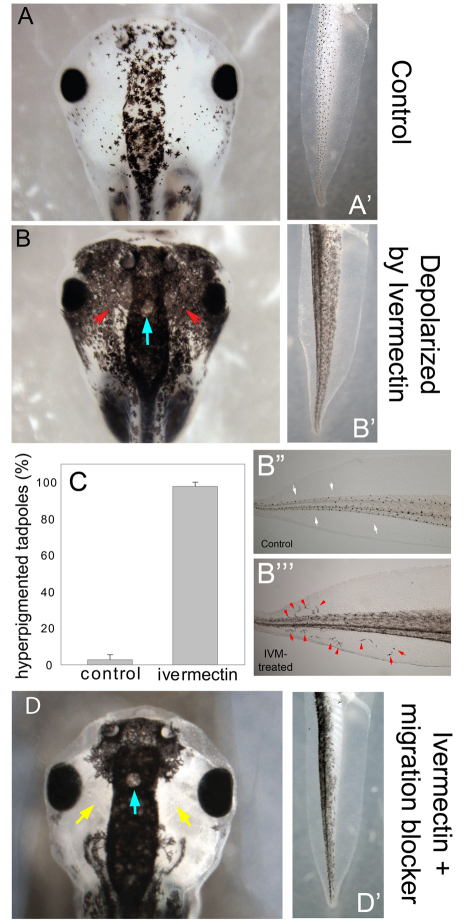
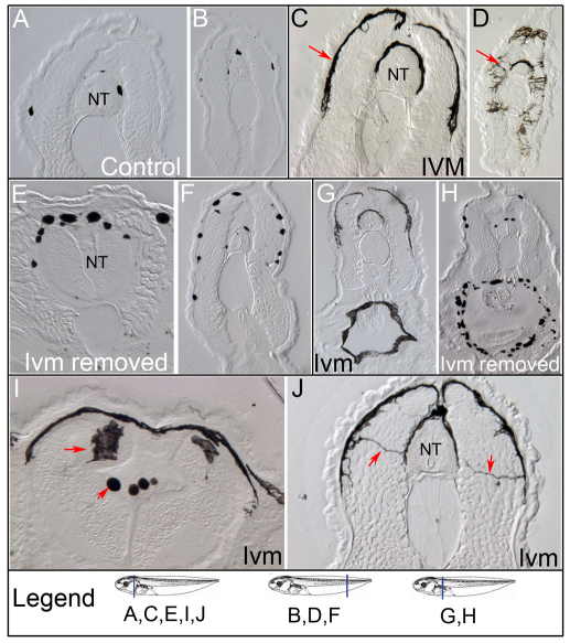
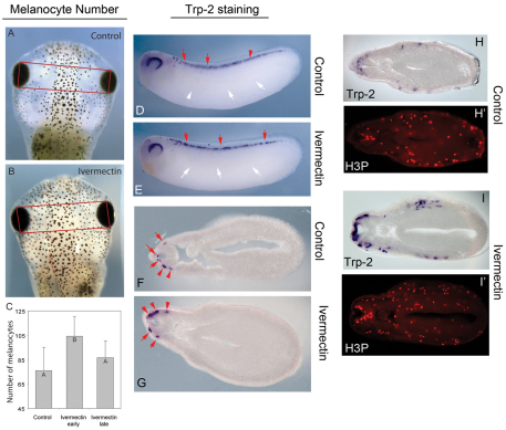
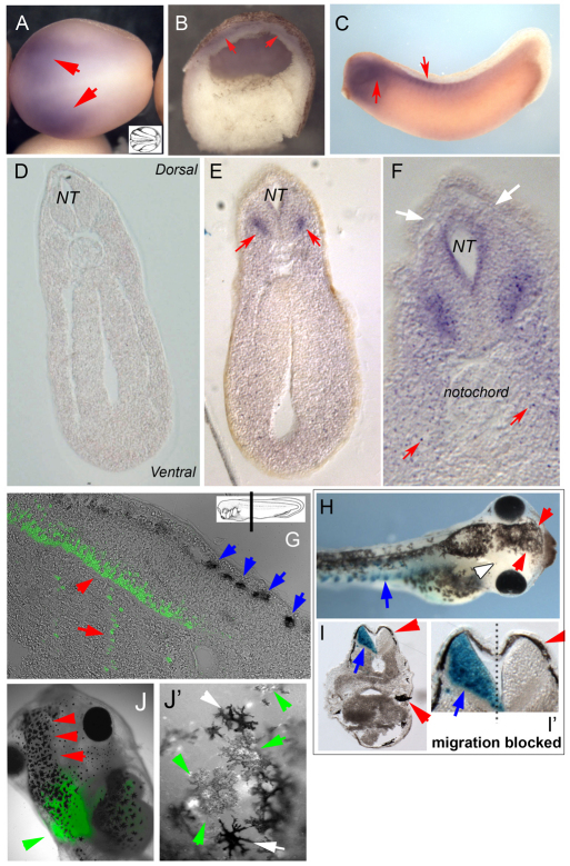
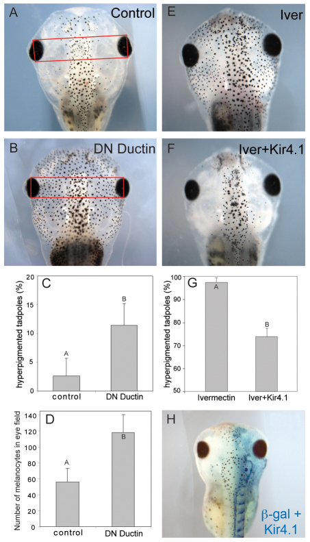
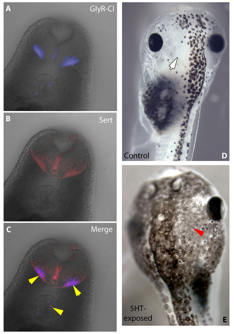
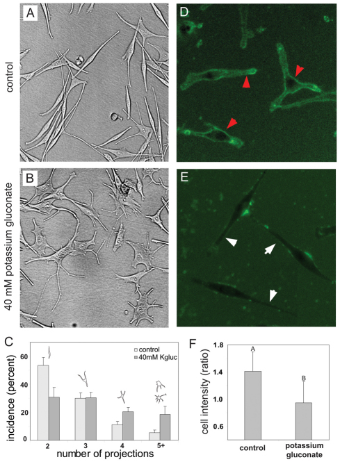
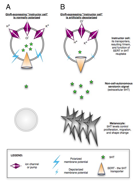
Understanding the mechanisms that coordinate stem cell behavior within the host is a high priority for developmental biology, regenerative medicine and oncology. Endogenous ion currents and voltage gradients function alongside biochemical cues during pattern formation and tumor suppression, but it is not known whether bioelectrical signals are involved in the control of stem cell progeny in vivo. We studied Xenopus laevis neural crest, an embryonic stem cell population that gives rise to many cell types, including melanocytes, and contributes to the morphogenesis of the face, heart and other complex structures. To investigate how depolarization of transmembrane potential of cells in the neural crest's environment influences its function in vivo, we manipulated the activity of the native glycine receptor chloride channel (GlyCl). Molecular-genetic depolarization of a sparse, widely distributed set of GlyCl-expressing cells non-cell-autonomously induces a neoplastic-like phenotype in melanocytes: they overproliferate, acquire an arborized cell shape and migrate inappropriately, colonizing numerous tissues in a metalloprotease-dependent fashion. A similar effect was observed in human melanocytes in culture. Depolarization of GlyCl-expressing cells induces these drastic changes in melanocyte behavior via a serotonin-transporter-dependent increase of extracellular serotonin (5-HT). These data reveal GlyCl as a molecular marker of a sparse and heretofore unknown cell population with the ability to specifically instruct neural crest derivatives, suggest transmembrane potential as a tractable signaling modality by which somatic cells can control stem cell behavior at considerable distance, identify a new biophysical aspect of the environment that confers a neoplastic-like phenotype upon stem cell progeny, reveal a pre-neural role for serotonin and its transporter, and suggest a novel strategy for manipulating stem cell behavior.


Figures

Fig. 1.
Fig. 1.
Ivermectin exposure induces hyperpigmentation. (A) Control embryos display a medially concentrated pigment pattern with the lateral eye field being largely devoid of melanocytes. (A′) The tail normally has a distributed population of round melanocytes over its core. (B) Embryos exposed to the chloride channel activator ivermectin while developing in the normal 10 mM Cl medium acquire a hyperpigmented phenotype by stage 42 despite otherwise normal development; ectopic melanocytes are present (periocular region indicated by red arrows; compare to similar region in panel A), and (B′) are more numerous and spread out in the tail. Ectopic melanocytes are also found in the dorsal and ventral fins (compare B‴ to control tails in B″). White arrows indicate fin region normally devoid of melanocytes; red arrowheads indicate ectopic melanocytes. (C) The ivermectin-induced phenotype was highly penetrant, with 98% of treated embryos developing hyperpigmentation (error bars indicate one standard deviation, n=189 for controls, n=174 for ivermectin-exposed). (D) When migration was blocked by the MMP inhibitor NSC-84093 in ivermectin-exposed embryos, colonization of ectopic locations by melanocytes was prevented (yellow arrows) but the abnormal arborization remained. The effect was also observed in the tail (D′), with the ventral area remaining uncolonized. Blue arrow indicates the location of an area that remains free of ectopic melanocytes, even in heavily hyperpigmented tadpoles, possibly overlying the pineal gland.
Fig. 2.
Fig. 2.
Ivermectin induces invasiveness in melanocytes. Compared to control embryos (A,B), those exposed to ivermectin (IVM) throughout development (C,D) show significantly more melanocyte coverage of the neural tube (NT) and epidermis (red arrows). Ivermectin-treated embryos removed from ivermectin at stage 43 (E,F) still show increased pigment cell number compared with controls, but the cells lose their arborized phenotypes. In addition, ivermectin induces melanocyte colonization of the gut (G,H) and the interior of the neural tube (I), and invasiveness of projections throughout the mesoderm between the epidermis and neural tube (J), indicated by red arrows. Schematics of Xenopus laevis embryo stages were retrieved from Xenbase, University of Calgary, Alberta T2N 1N4, Canada; http://www.xenbase.org/; August 2010.
Fig. 3.
Fig. 3.
Early ivermectin exposure induces an increase in pigment cell proliferation. Embryos exposed to ivermectin from stages 10–24 (early) or 28–46 (late) both show darkening due to expansion of melanocytes. To determine whether there was also a corresponding increase in melanocyte number, photographs were taken of controls (A) and ivermectin-exposed (B) embryos after tricaine anesthetization, which contracts the pigment cells. The number of melanocytes in the eye field (red boxes) were then counted. Early exposed embryos showed a 1.5-fold increase in melanocyte number relative to controls (C), whereas no detectable difference was observed between late exposed embryos and controls. Error bars indicate one standard deviation; n=24 embryos for each treatment. Control embryos processed in in situ hybridization for the melanocyte marker Trp2 at stage 28 show the normal pattern of expression prior to the migration of melanocytes away from the dorsal neural tube (D). Ivermectin-treated embryos show precisely the same pattern (E) and exhibit no evidence of ectopic locations being converted into a melanocyte fate by the ivermectin treatment. Sectioning reveals that control (F) and ivermectin-treated (G) embryos have the same number of melanocytes at the neural tube, also ruling out local shifts of neural crest cells into the melanocyte lineage as the explanation for later hyperpigmentation. Red arrows indicate positive signal (melanocytes indicated by Trp2 expression), whereas white arrows indicate lack of signal. (H-I′) To directly analyze proliferation in melanocytes, embryos were stained for the melanocyte marker Trp2 using in situ hybridization to identify pigment cells, and were then sectioned and processed for immunohistochemistry with anti-H3B-P antibody. (H) Trp2 section in control embryos; (H′) corresponding signal of H3B-P stain in the same section. (I) Trp2 section in ivermectin-exposed embryos; (I′) corresponding signal of H3B-P stain in the same section. Overlays of the bright-field and fluorescent signals from the same section allowed quantification of the number of melanocytes that were in mitosis. At stage 28, there was no difference (P>0.2, n=6) between controls and ivermectin-treated embryos. By stage 35, there was a significant increase in the number of mitotic melanocytes in the ivermectin-treated embryos (P<0.009, n=6).
Fig. 4.
Fig. 4.
Expression of GlyCl-α mRNA and protein. In situ hybridization was performed on Xenopus embryos with an antisense probe to GlyCl-α. Expression (red arrows) was first detected during neurulation in the developing neural plate (A; panel B shows a thick section in profile because expression was too weak to be clearly visible in thin sections). Expression became restricted during somite stages (C) with foci of staining observed in the ventral marginal zone of the neural tube (NT) (E). Sections also revealed punctate signal in the lateral mesoderm (F, red arrows), which was not observed in the no-primary control (D), and an absence of signal in the dorsal neural tube, where many melanocytes are located (F, white arrows). In panels A–F, red arrows indicate expression of GlyCl mRNA, whereas white arrows indicate lack of expression in the dorsal neural tube from which melanocytes originate. Immunohistochemistry (with an antibody to GlyCl; green signal and red arrowheads) and in situ hybridization (with a probe to the melanocyte marker Trp2, blue arrows) on the same section of stage-31 embryos (G) revealed that the cells expressing GlyCl are at some distance from melanocytes (melanocytes do not themselves express the ivermectin target protein). (H) As an additional test of long-range signaling, embryos were injected with KCNE1+β-gal mRNA at the 16-cell stage in blastomeres, which resulted in depolarizing potassium channel subunit expression in posterior ventral tissues (blue arrow indicates β-galactosidase lineage label). Red arrowheads in panels H,I indicate hyperpigmentation (aberrant melanocytes) in the region; white arrowhead in H indicates absence of β-gal signal from anterior regions. They were continuously treated with NSC-84093 to prevent melanocyte migration from distant regions of the embryo. Sectioning (I,I′) revealed that hyperpigmentation occurred in the head and on the contralateral side, demonstrating that the metastasis-inducing signal is able to cross considerable distance along the anterior-posterior axis (from somites over the gut into the space anterior to the eyes) and across the embryonic midline (red arrowhead in I′) even when melanocytes local to the KCNE1 depolarization are prevented from moving. Insets in A and G taken from Nieuwkoop and Faber (Nieuwkoop and Faber, 1967); schematic inset in G shows plane of sections for panels D–G. (J) A small section of neural plate from a ubiquitous GFP-transgenic donor treated with ivermectin (green arrowheads) was transplanted into an untreated host at stage 18, resulting in a hyperpigmentation phenotype (red arrowheads). (J′) Similar transplant performed from an ivermectin-treated donor results in GFP-labeled melanocytes (lighter in color owing to overlap of fluorescence and black pigment; green arrowheads) shows that these melanocytes take up ectopic positions next to native melanocytes (white arrowheads) and acquire the same highly arborized shape.
Fig. 5.
Fig. 5.
Hyperpigmentation is due to depolarization. Microinjection of a dominant-negative form of ductin (dn-xDuct) at the one-cell stage inhibits the hyperpolarizing H+-V-ATPase and results in hyperpigmentation (A,B). Injections result in hyperpigmented tadpoles in 11.5% of embryos (C), significantly higher than background levels observed in control embryos. Hyperpigmented embryos arising from dn-xDuct injections were photographed and the number of melanocytes in the eye field counted; there was a 2.1-fold increase in number of melanocytes compared with age-matched controls (D). By contrast, overexpression of the hyperpolarizing potassium channel Kir4.1 (E,F) inhibits ivermectin-induced hyperpigmentation in 25% of injected embryos (G). Kir4.1-mediated inhibition was non-cell-autonomous, because one of two cell injections, resulting in hyperpolarizing channel activity on just one side of the embryo, inhibited hyperpigmentation on both the left and right side of the embryos (H).
Fig. 6.
Fig. 6.
Serotonergic controls of melanocyte behavior and their relationship to GlyCl-expressing cells. (A) Sections of a stage 32 embryo processed by immunohistochemistry with an anti-GlyCl antibody and visualized with a fluorescent secondary antibody (Alexa Fluor 647). (B) The same section processed by immunohistochemistry with an anti-SERT antibody and visualized with Alexa Fluor 546. (C) Merge of A and B showing colocalization of GlyCl and SERT. Yellow arrowheads indicate areas of overlapping expression (cells containing both GlyCl and SERT). Unlike controls (D), embryos treated with external serotonin acquire the hyperpigmentation phenotype (E), consistent with SERT mediating the effect of depolarized GlyCl-expressing cells on melanocytes. White arrow indicates a region normally devoid of melanocytes; red arrowhead indicates ectopic melanocytes.
Fig. 7.
Fig. 7.
Human melanocytes exhibit arborization when the membrane is depolarized. In normal culture medium, human melanocytes typically develop two or three projections (A). When grown in media supplemented with 50 mM potassium gluconate, cells develop a more arborized morphology, with many cells having four or five, or more, projections (B). Comparisons between treatments (C) demonstrate a significant effect of potassium gluconate on arborization of melanocytes. Error bars indicate one standard deviation. Image analysis (using the membrane voltage sensor pair CC2-DMPE and DiBAC4) comparing controls (D) and cells cultured in high-potassium medium (E) revealed the predicted depolarization (lower intensity of pixels) in response to the high-potassium media (F). Red arrowheads indicate depolarized cell membranes; white arrows indicate lack of depolarization in membrane.
Fig. 8.
Fig. 8.
A model of melanocyte control by transmembrane potential of cells in the neural crest’s environment. (A) In unperturbed embryos, several classes of ion transporters keep the plasma membrane polarized. This transmembrane potential powers the reuptake of extracellular serotonin through its transporter SERT, resulting in normal melanocyte behavior. (B) By contrast, when the instructor cell population (demarcated by GlyCl expression) is depolarized by targeted modulation of H+, Cl or K+ channel/pump function, the SERT runs backwards and not only fails to clear the extracellular space of serotonin, but actually exports additional serotonin. The higher serotonin level in the milieu of the neoblasts induces neoplastic-like behavior in melanocytes, as occurs in human cancers. This pathway can be manipulated at a number of points. Consistent with this model, our data show that, although direct serotonin exposure or depolarization of GlyCl-expressing cells can induce hyperpigmentation, the depolarization phenotype can be prevented by overexpression of hyperpolarizing channels or inhibition of SERT. Central features of this model are the regulation of cell behavior by transmembrane potential, regardless of which specific gene product achieves it, and non-cell-autonomous effects of a cell subpopulation specifically instructing, at considerable distance, one derivative of neural crest to undergo the stem-cell-to-neoplastic-cell-like phenotype.

References

    1. Aberg P., Geladi P., Nicander I., Hansson J., Holmgren U., Ollmar S. (2005). Non-invasive and microinvasive electrical impedance spectra of skin cancer-a comparison between two techniques. Skin Res. Technol. 11, 281–286 - PubMed
    1. Adams D.S. (2008). A new tool for tissue engineers: ions as regulators of morphogenesis during development and regeneration. Tissue Eng. Part A 14, 1461–1468 - PubMed
    1. Adams D.S., Levin M. (2006a). Inverse drug screens: a rapid and inexpensive method for implicating molecular targets. Genesis 44, 530–540 - PMC - PubMed
    1. Adams D.S., Levin M. (2006b). Strategies and techniques for investigation of biophysical signals in patterning. In Analysis of Growth Factor Signaling in Embryos (ed. Whitman M., Sater A.K.), pp. 177–262 Taylor and Francis Books
    1. Adams D.S., Robinson K.R., Fukumoto T., Yuan S., Albertson R.C., Yelick P., Kuo L., McSweeney M., Levin M. (2006). Early, H+-V-ATPase-dependent proton flux is necessary for consistent left-right patterning of non-mammalian vertebrates. Development 133, 1657–1671 - PMC - PubMed

Publication types

MeSH terms