. 2011 Jan;4(1):86-94.
doi: 10.1242/dmm.005777. Epub 2010 Oct 19.

Enterohemorrhagic Escherichia coli induce attaching and effacing lesions and hemorrhagic colitis in human and bovine intestinal xenograft models

Affiliations

Enterohemorrhagic Escherichia coli induce attaching and effacing lesions and hemorrhagic colitis in human and bovine intestinal xenograft models

Lilach Golan et al. Dis Model Mech. 2011 Jan.

Abstract

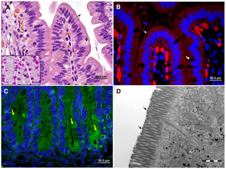
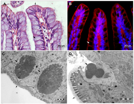
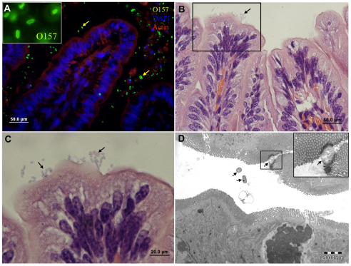
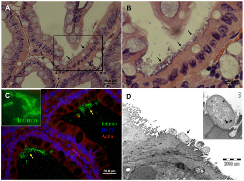
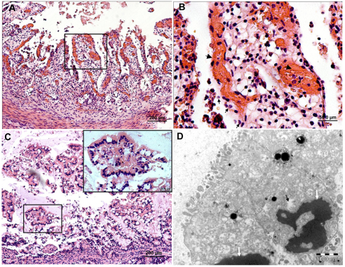
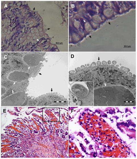
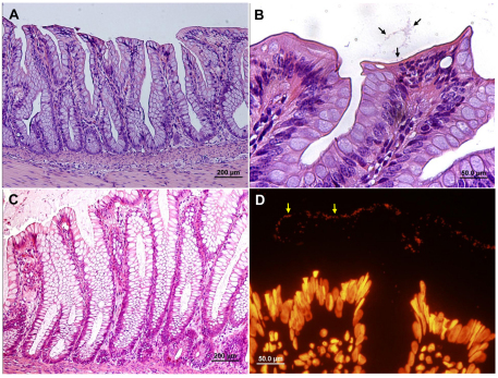
Enterohemorrhagic Escherichia coli (EHEC) O157:H7 is an important cause of diarrhea, hemorrhagic colitis and hemolytic uremic syndrome in humans worldwide. The two major virulence determinants of EHEC are the Shiga toxins (Stx) and the type III secretion system (T3SS), including the injected effectors. Lack of a good model system hinders the study of EHEC virulence. Here, we investigated whether bovine and human intestinal xenografts in SCID mice can be useful for studying EHEC and host tissue interactions. Fully developed, germ-free human and bovine small intestine and colon were established by subcutaneous transplantation of human and bovine fetal gut into SCID mice. Xenografts were allowed to develop for 3-4 months and thereafter were infected by direct intraluminal inoculation of Stx-negative derivatives of EHEC O157:H7, strain EDL933. The small intestine and colon xenografts closely mimicked the respective native tissues. Upon infection, EHEC induced formation of typical attaching and effacing lesions and tissue damage that resembled hemorrhagic colitis in colon xenografts. By contrast, xenografts infected with an EHEC mutant deficient in T3SS remained undamaged. Furthermore, EHEC did not attach to or damage the epithelium of small intestinal tissue, and these xenografts remained intact. EHEC damaged the colon in a T3SS-dependent manner, and this model is therefore useful for studying the molecular details of EHEC interactions with live human and bovine intestinal tissue. Furthermore, we demonstrate that Stx and gut microflora are not essential for EHEC virulence in the human gut.


Figures

Fig. 1.
Fig. 1.
Bovine fetal small intestinal xenograft in SCID mice. Representative microscopic images of fully differentiated small intestinal xenografts. (A) H&E and PAS staining of formalin-fixed paraffin sections. (B,C) Fluorescence staining of cryosections with phalloidin-rhodamine (red, B) or anti-cytokeratin antibodies (green, C) combined with DAPI (blue). (D) TEM image of xenograft. Normal, fully developed villous (A,B) and crypt (C) mucosal epithelia are visible. Microvilli are clearly visible on the apical (luminal) border of the villous epithelial cells (A,B,D; small arrows), which are interspaced with a small number of goblet cells (A,B; large arrows) containing PAS-positive mucus (inset A; arrow). Crypts of the small intestinal mucosa with numerous Paneth cells (arrows) are shown in C. Scale bars: 50 μm (A-C); 1 μm (D).
Fig. 2.
Fig. 2.
Bovine fetal colon xenograft in SCID mice. Representative microscopic images of H&E-stained formalin-fixed paraffin sections (A), fluorescence staining of cryosections with phalloidin-rhodamine (red) combined with DAPI (blue) (B) and TEM (C, D) of fully differentiated colon xenografts. Normal, fully developed colonic mucosa are composed of numerous goblet cells (large arrows) containing characteristic mucus granules (C,D) which are excreted through the luminal orifice of the cell (D). Microvilli are visible on the apical (luminal border) of the epithelial cells (A,C,D; small arrows) and are covered by the glycocalyx layer (D; small arrow). Scale bars: 50 μm (A,B); 2 μm (C); 1 μm (D).
Fig. 3.
Fig. 3.
EHEC are not pathogenic in the bovine small intestine and only sparsely adhere to the villous epithelium. Representative microscopic images of tissue sections of bovine small intestinal xenografts in SCID mice 8 hours after intraluminal challenge with 4×10⁷ organisms. Xenograft cryosection stained with anti-O157 antibody (green) combined with DAPI (blue) and phalloidin-rhodamine (red) (A), H&E stained formalin-fixed sections (B,C) and TEM image (D). Mucosal epithelium is intact and undamaged and most organisms are not attached to the mucosal epithelium (arrows in A and D), whereas adhering organisms (arrows in C, and D inset) do not induce the characteristic AE lesions. Scale bars: 50 μm (A,B); 20 μm (C); 2 μm (D).
Fig. 4.
Fig. 4.
EHEC induce formation of AE lesions on bovine colon epithelial cells. Representative microscopic images of tissue sections of bovine colon xenografts in SCID mice 8 hours after intraluminal challenge with 4×10⁷ organisms. H&E staining of formalin-fixed paraffin sections (A,B), fluorescence staining of cryosections with anti-intimin antibody (green) combined with DAPI (blue) and phalloidin-rhodamine (red) (C) and TEM (D). EHEC adhere to mucosal epithelial cells (A-C; arrows) expressing intimin (C and inset image) and induce formation of AE lesions (D). Pedestal formation under adhering bacterial cell (arrow) is demonstrated (D) and better seen in the enlarged inset image. Scale bars:50 μm (A,C); 20 μm (B); 2 μm (D).
Fig. 5.
Fig. 5.
Hemorrhagic colitis and epithelial damage induced by EHEC infection of the bovine colon. Microscopic images of tissue sections of bovine colon xenografts in SCID mice 8 hours after intraluminal challenge with 4×10⁷ organisms. H&E staining of formalin-fixed paraffin sections (A–C) and TEM (D). Epithelial damage associated with hemorrhages (B, large arrows) and leukocyte infiltration (B, small arrows). In other infected colon xenografts, epithelial damage is not associated with signs of inflammation (C) and cells appear with condensed pyknotic nuclei, damaged membranes and cytoplasmic vacuolization (D, arrows). Scale bars: 200 μm (A,C); 50 μm (B); 1 μm (D).
Fig. 6.
Fig. 6.
EHEC infecting human colon induce formation of AE lesions and tissue damage. Representative microscopic images of tissue sections of human colon xenografts in SCID mice 8 hours after intraluminal challenge with 4×10⁷ organisms. H&E staining of formalin-fixed paraffin sections (A,B,E,F) and TEM (C,D). Bacteria adhere to mucosal epithelial cells (black arrows in A and B) and induce formation of AE lesions (B). AE lesions and pedestal formation under adhering bacterial cells (black arrows) are demonstrated (C) and better seen in the enlarged image (D and inset image). Goblet cells are indicated by white arrows in all images. Scale bars: 50 μm (A); 20 μm (B); 5000 nm (C); 2000 nm (D). Some colon segments showed severe epithelial damage and hemorrhages only sparsely infiltrated by leukocytes (E) and this is better seen in the boxed area enlarged in F. Scale bars: 200 μm (E); 50 μm (F).
Fig. 7.
Fig. 7.
EHEC-induced formation of AE lesions and tissue damage are T3SS-dependent processes. Representative microscopic images of tissue sections of bovine (A,B) and human (C,D) colon xenografts in SCID mice 8 hours after intraluminal challenge with 4×10⁷ EHEC of a strain with deleted escN gene (encoding a T3SS-associated ATPase), which is unable to assemble a functional T3SS. H&E staining of formalin-fixed paraffin sections (A–C) and fluorescence staining of cryosections with Sytox Orange staining for nucleic acids (D). The bovine (A,B) and human (C,D) colon mucosa remained unaffected, showing normal mucosal architecture and nonadhering organisms in the lumen (B and D, arrows). Scale bars: 200 μm (A,C); 50 μm (B,D).

Similar articles

Cited by

References

    1. Cantey J.R., Inman L.R., Blake R.K. (1989). Production of diarrhea in the rabbit by a mutant of Escherichia coli (RDEC-1) that does not express adherence (AF/R1) pili. J. Infect. Dis. 160, 136–141 - PubMed
    1. Chase-Topping M., Gally D., Low C., Matthews L., Woolhouse M. (2008). Super-shedding and the link between human infection and livestock carriage of Escherichia coli O157. Nat. Rev. Microbiol. 6, 904. - PMC - PubMed
    1. Chong Y., Fitzhenry R., Heuschkel R., Torrente F., Frankel G., Phillips A.D. (2007). Human intestinal tissue tropism in Escherichia coli O157: H7-initial colonization of terminal ileum and Peyer’s patches and minimal colonic adhesion ex vivo. Microbiology 153, 794–802 - PubMed
    1. Croxen M.A., Finlay B.B. (2010). Molecular mechanisms of Escherichia coli pathogenicity. Nat. Rev. Microbiol. 8, 26–38 - PubMed
    1. Fitzhenry R.J., Pickard D.J., Hartland E.L., Reece S., Dougan G., Phillips A.D., Frankel G. (2002). Intimin type influences the site of human intestinal mucosal colonisation by enterohaemorrhagic Escherichia coli O157:H7. Gut 50, 180–185 - PMC - PubMed

Publication types

MeSH terms

Substances