. 2010 Oct 19:6:423.
doi: 10.1038/msb.2010.80.

Molecular characterization of the evolution of phagosomes

Affiliations

Molecular characterization of the evolution of phagosomes

Jonathan Boulais et al. Mol Syst Biol. .

Abstract

Amoeba use phagocytosis to internalize bacteria as a source of nutrients, whereas multicellular organisms utilize this process as a defense mechanism to kill microbes and, in vertebrates, initiate a sustained immune response. By using a large-scale approach to identify and compare the proteome and phosphoproteome of phagosomes isolated from distant organisms, and by comparative analysis over 39 taxa, we identified an 'ancient' core of phagosomal proteins around which the immune functions of this organelle have likely organized. Our data indicate that a larger proportion of the phagosome proteome, compared with the whole cell proteome, has been acquired through gene duplication at a period coinciding with the emergence of innate and adaptive immunity. Our study also characterizes in detail the acquisition of novel proteins and the significant remodeling of the phagosome phosphoproteome that contributed to modify the core constituents of this organelle in evolution. Our work thus provides the first thorough analysis of the changes that enabled the transformation of the phagosome from a phagotrophic compartment into an organelle fully competent for antigen presentation.


Conflict of interest statement

The authors declare that they have no conflict of interest.

Figures

Figure 1
Figure 1
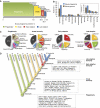
Shared components define the ‘ancient’ phagosome. (A) Predicted orthologs of phagosome proteins of Dictyostelium, Drosophila, and mouse were analyzed by BLAST against the two other species and mapped according to −Log10(e-value), where 0 indicates the absence of an ortholog and 181 a perfect alignment. Four distinct groups of proteins are highlighted for each organism: (1) a set of orthologs shared by the three organisms defining the ‘ancient’ phagosome (blue data points outside the x and y axes), (2 and 3) groups of conserved proteins shared only between the plotted organism and one of the two others found on the x or y axis (green or red data points), and (4) a set of proteins unique to the plotted organism (purple data points at the origin of the graph). As several data points may overlay in the scatter plot, a histogram below each plot reports the relative distribution of proteins among the four distinct groups of proteins. (B) Annotation of a function to each protein of the mouse proteome highlights the level of conservation of relevant phagosome functions among the three organisms. Although a large proportion of the proteins associated with functions such as ‘membrane trafficking,’ ‘small GTPases,’ and ‘cytoskeleton’ are majorly shared by the three organisms, some like ‘membrane receptors’ and ‘immunity’ are more specific to mouse and Drosophila phagosomes. See also Supplementary Figure S1 and Supplementary Datasets 4–6.
Figure 2
Figure 2
Origin of the mouse phagosome proteome. Comparative analyses of the mouse phagosome proteome among 39 taxa identified the origin of each protein. (A) Proportions (in %) of the evolutionary origin of the mouse phagosome proteome are reported through four major evolutionary groups of proteins: phagotrophy (Eukaryota, Amoebozoa, and Fungi), innate immunity (Bilateria, Coelomata, and Chordata), early (Euteleostomi), and late adaptive immunity (Tetrapoda and beyond). (B) Comparison between the evolutionary origin of the mouse phagosome proteins and the entire mouse proteome (reported by their relative proteome proportion in %) through a cladistic distribution (x axis) reveals that phagosomes are of ancient origin. The inbound graph shows the same proteome proportion in % through a cladistic distribution under the four major evolutionary groups of proteins reported in a: phagotrophy (Ph.), innate immunity (In.), early adaptive (E.A.), and late adaptive immunity (L.A.). (C) Comparative functional analysis of the mouse phagosome proteins reveals that specific phagosomal functions originated from different stages of evolution. The function ‘Others’ contains the merging of remaining functions, and numbers indicate the amount of proteins found in each function. (D) Specific examples of proteins originating at the four major evolutionary groups are found in dash boxes. See also Supplementary Dataset 7.
Figure 3
Figure 3
Novel components of the mouse phagosome emerged through two major periods of gene duplication. (A) Of 952 pairs of duplicated genes encoding for mouse phagosomal proteins, the majority of these genes have their origin in the phagotrophy stage of evolution. (B) Proportional representation of the origin of gene duplication events on the phagosome and the whole mouse genome shows that duplication of genes encoding mouse phagosomal proteins occurred mainly in Bilateria (emergence of innate immunity) and Euteleostomi (emergence of adaptive immunity), whereas gene duplication in the whole murine genome occurred more evenly throughout evolution. (C) Functional analysis of phagosomal proteins duplicated originally in bilaterians and euteleosts reveals a preference for small GTPases, signaling, and proteins involved in cellular trafficking. The function ‘Others’ contains the merging of remaining functions. See also Supplementary Figure S3 and Supplementary Dataset 8.
Figure 4
Figure 4
Evolution of the phagosome phosphoproteome. (A) Alignment of mouse phagosomal phosphoproteins revealed strong conservation of phosphosites within mammals, but fewer phosphosites are conserved across vertebrates, chordates, and tetrapods. In proportion, a larger fraction of conserved phosphosites (in red) is observed in ordered regions compared with disordered regions. (B) Phosphosites modulated by IFN-γ are on average as conserved as other phosphorylated residues in vertebrates but not in tunicates or Drosophila. (C) Comparative alignment of conserved mouse, Drosophila, and Dictyostelium phagosomal phosphoproteins identified by MS revealed that the majority (near 66%—doughnut plot) of phosphosites are not conserved (blue), indicating that the mouse phagosome phosphoproteome is globally recent in evolution. Still, around 33% of mouse phosphosites are phosphorylatable (red) in Drosophila or Dictyostelium. Of these, 12.8 and 5.0% phosphosites were observed to be phosphorylated (green) in Drosophila and Dictyostelium phagosomes, respectively (Obs.). These phosphosites are 8 and 12 times more conserved compared with random S/T/Y (expected, Exp.) of the same phosphoproteins in Drosophila and Dictyostelium, respectively. Bars indicate 95% confidence interval.
Figure 5
Figure 5
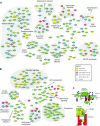
Evolution of the phagosome proteins network. Experimental data from the Intact database and curated entries of the UniProt database were used to generate a network from protein–protein interactions of identified mouse phagosomal proteins. From the total network, subnetworks of cytoskeleton and vesicle trafficking proteins (A) and immunity-related proteins (B) were extracted, showing the evolutionary mixed origin of most protein complexes and the addition of novel modules such as the MHC class I and II presentation machinery, the receptor signaling, the NADPH oxidase complex or the Ena/VASP complex to the phagosome in evolutionary steps of adaptive and innate immunity, respectively. Duplicated proteins of which both paralogs have been identified by MS/MS on mouse phagosomes are circled in blue. (C) Example how duplication might affect phagosome function: (immuno-) proteasome activator complex subunits PSME1 (PA28α) and PSME2 (PA28β) were duplicated with the appearance of jawed fishes, coinciding with the emergence of adaptive immunity. During this duplication event, PSME1 gained a KEKE-motif that was not present in the common ancestor. KEKE-motifs have been described to interact with each other and are also present in several chaperones including Calnexin (Li and Rechsteiner, 2001; Rechsteiner and Hill, 2005). It is likely that introduction of the KEKE-motif in PSME1 might locate the immunoproteasome to Calnexin and the MHC class I presentation machinery, thereby enhancing antigen presentation efficiency.
Figure 6
Figure 6
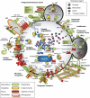
Role of molecular machines of mixed origin in phagosome functions. Many of the functional properties of mammalian phagosomes involve molecular machines made of proteins that emerged at different periods during evolution. For example, in the context of antigen cross-presentation, key steps such as phagosome/endosome fusion, the killing of microbes and their degradation into peptides, as well as their loading on MHC class I molecules are made possible by proteins that appeared in organisms where the phagosome has its main role in phagotrophy (green proteins), innate immunity (yellow proteins), and adaptive immunity (red proteins). Remarkably, the emergence of a cytokine such as IFN-γ inteleosts, >1.2 billion years after the emergence of phagotrophy (Bhattacharya et al, 2009), allowed the fine-tuning of the expression and/or phosphorylation of proteins of each of these groups (red shadow). Early endosome (EE), late endosome (LE), lysosome (Ly), and endoplasmic reticulum (ER).

References

    1. Ackerman AL, Kyritsis C, Tampe R, Cresswell P (2003) Early phagosomes in dendritic cells form a cellular compartment sufficient for cross presentation of exogenous antigens. Proc Natl Acad Sci USA 100: 12889–12894 - PMC - PubMed
    1. Altschul SF, Madden TL, Schaffer AA, Zhang J, Zhang Z, Miller W, Lipman DJ (1997) Gapped BLAST and PSI-BLAST: a new generation of protein database search programs. Nucleic Acids Res 25: 3389–3402 - PMC - PubMed
    1. Barabasi AL, Oltvai ZN (2004) Network biology: understanding the cell′s functional organization. Nat Rev Genet 5: 101–113 - PubMed
    1. Berglund AC, Sjolund E, Ostlund G, Sonnhammer EL (2008) InParanoid 6: eukaryotic ortholog clusters with inparalogs. Nucleic Acids Res 36: D263–D266 - PMC - PubMed
    1. Bertholet S, Goldszmid R, Morrot A, Debrabant A, Afrin F, Collazo-Custodio C, Houde M, Desjardins M, Sher A, Sacks D (2006) Leishmania antigens are presented to CD8+ T cells by a transporter associated with antigen processing-independent pathway in vitro and in vivo. J Immunol 177: 3525–3533 - PubMed

Publication types

LinkOut - more resources